News


22 July 2026
Coral Keepers
Inside our coral spawning success. In the heart of the midwest.
Read Article
News
Unless you’ve had cancer or supported someone who has, you may not be aware of the Herculean efforts often required to battle the disease. But if you are a cancer survivor, you’ve likely thanked an oncologist, possibly even the nurses and technicians at your cancer hospital.
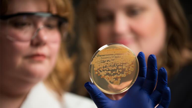

By Elise Lamar, PhD
Unless you’ve had cancer or supported someone who has, you may not be aware of the Herculean efforts often required to battle the disease. But if you are a cancer survivor, you’ve likely thanked an oncologist, possibly even the nurses and technicians at your cancer hospital. Equally worthy of gratitude are the donors whose names are on the building and the fundraisers whose cancer awareness events raise research dollars. Then there are the drug company researchers who turn molecules into cancer therapies as well as the volunteers who undergo clinical trials to optimize these products. All work to fight this insidious disease.
And there are still more people who may have had a hand in a positive outcome. Yet, you probably didn’t meet them at the hospital, in your oncologist’s office, or at a fundraiser, as they’re almost always working, preferring to avoid the limelight. But these driven individuals may have played a big part in your recovery.
They are basic research scientists.
A matter of intent
Associate Investigator Sue Jaspersen, PhD, personifies the curiosity-driven mindset of basic scientists at the Stowers Institute for Medical Research. For almost two decades she has studied the biochemical nuts and bolts of cell division in yeast. Her goal? “To understand how cells make decisions about how to grow, divide, make copies of their DNA, and distribute it to daughter cells so that they are healthy and happy and have everything they need to make healthy and happy children of their own!”
Missing from Jaspersen’s list is the intention to cure a disease or develop a pharmaceutical. Jaspersen, also an associate professor in the Department of Molecular and Integrative Physiology at the University of Kansas School of Medicine, simply wants to know how cells divide and thinks yeast is the best organism for studying this process. Even without a direct, cure-related purpose, the American Cancer Society awarded her a grant in 2011 to study proteins that sit on the inner face of a yeast cell nuclear membrane: “because that area interacts with chromosomes, and knowing how proteins get to this space might tell us how chromosomes stay organized.”
Keeping chromosomes organized is critical. A cancer’s signature is chromosomal chaos, or what biologists call genomic instability. Marked by damage such as mutations in DNA strands or abnormal numbers of chromosomes, this genomic instability causes uncontrolled cell division, the trait all cancers share.
Jaspersen’s research does not directly address how to avert genomic disaster, but it has unmistakable relevance to cancer, as does any biochemical analysis of cell division. “Understanding how cells do something right will eventually lead to knowing how to treat cancers or inherited diseases in which normal cell division is subverted,” says Jaspersen.
One of her current interests is how multiprotein complexes called spindle pole bodies (SPBs) in yeast duplicate themselves one time in preparation for cell division. That event kicks off construction of a gigantic molecular scaffold called the mitotic spindle anchored at each end by SPBs. In a process Jaspersen likens to tugging on a wishbone, replicated chromosomes then get dragged in opposite directions by the spindle into those healthy and happy (if the wishbone is perfectly bisected) daughter cells.
Jaspersen’s lab wants to identify molecules that orchestrate this event. For example, in a 2014 collaborative study published in PLoS Genetics, Jaspersen and colleagues showed how one protein in the crowd of SPB proteins controlled whether the entire complex duplicated. This work has potential applications to human health, as cancer cells often contain more than one SPB (called centrosomes in mammals), a mistake that might be linked to genomic breakdown.
She says obtaining funding to conduct pure research like this can be challenging because many donors want to see a near-term impact of their philanthropy. “But what is not always clear,” she says, “is that funding basic research is a proven route to new and better ways of treating disease.”
“You’re the scientist.”
The idea is not controversial at the Institute. In fact, it is why Jim and Virginia Stowers created the Institute. As an illustration, Associate Investigator Tatjana Piotrowski, PhD, recalls a conversation she had with Jim Stowers in 2010. When asked what he might like her to work on, Mr. Stowers said, “Well, you’re the scientist. You should know what is important."
What Piotrowoski deemed important was understanding how cells move. So she built her laboratory around zebrafish as a model organism and decided to study development of sensory hairs that fish use to detect water movement. This sense organ, called the lateral line, forms as a procession of immature precursor cells march collectively from the head to the tail of a fish and mature in a line as they go. But since only aquatic vertebrates have a lateral line, why should a mammal care?
For numerous reasons, not the least being that injured lateral line cells regenerate. Figuring out how they do that, and why analogous hair cells in the human inner ear cannot, might suggest ways to reverse some forms of hearing loss in humans. But embryonic lateral line cells also share the second worst attribute of cancer cells - they migrate in a manner reminiscent of metastatic cancer cells that travel in packs. “If we want to study collective cell migration in a living animal, we must use model organisms, like fish,” says Piotrowski. “It is not an exaggeration that a migrating fish cell likely uses cues similar to those used by migrating human cells.”
Findings from the Piotrowski lab published this year in Cell Reports drive that point home. To define signals that guide lateral line precursors, the researchers first employed genetic techniques to delete a large protein displayed on the membrane of those cells called heparan sulfate proteoglycan (HSPG) and then tracked the cells’ progress. HSPG loss caused cells to tumble chaotically rather than move forward in a disciplined fashion. Antennae-like HSPGs are often decorated with complex sugars, or glycans. The work suggests that environmental cues transduced by these glycosylated membrane proteins keep cells marching in line.
Interestingly, HSPGs also extend from certain human cancer cells, among them metastatic breast cancers and melanoma, and clinicians correlate changes in their structure with unfavorable prognosis. Drugs that control HSPG synthesis, called heparanase inhibitors, are being evaluated as anticancer drugs in clinical trials. Whether metastatic human tumor cells use the same signals employed by fish lateral line precursors remains unknown. But if they do, Piotrowski suggests that candidate antimetastatic drugs could be developed using knowledge gained from studying the fish.
Not something a cancer cell invented
Developmental biologist Paul Kulesa, PhD, who directs the Institute’s Imaging Center, also studies migration of motile embryonic cells - in his case, neural crest cells in a chick embryo model system. In vertebrates, neural crest cells fan out from the head and embryonic spinal cord to form diverse structures such as facial features, smooth muscle, pigmented cells, and the peripheral nervous system. Like Piotrowski’s zebrafish, chick embryos are an ideal system because you can follow a single cell in a living animal.
Jennifer Kasemeier-Kulesa, a research specialist II in the lab, notes that invasiveness is not a sinister attribute but rather a necessity for many well-behaved cells in a developing embryo. “Getting from one point to another in an organism is not something a cancer cell invented,” she says. “Cancer cells migrate by pre-empting signals used by normal cells.”
Proof for that comes from the lab’s ongoing analysis of two neural crest-derived cancers: melanoma, a cancer of pigmented cells, and neuroblastoma, a malignancy of sympathetic nervous system precursors. In a study published in Pigment Cell & Melanoma Research in 2012, Kulesa and colleagues reported that human melanoma cells apparently “remember” that they were once neural crest cells, because they migrate along neural crest pathways when placed in a chick embryo. This may mean that tumor cells retain the molecular detection gear—analogous to Piotrowski’s HSPG proteins—required to pick up guidance signals emitted in their ancestral neighborhood.
The Kulesa lab recently received a grant from the National Institutes of Health (NIH) to determine if human neuroblastoma cells behave similarly; they’re optimistic, based on their recent discovery of a signaling pathway that controls migration of normal sympathetic nervous system precursors.
Neuroblastoma is the most common cancer of infants. Children born with the disease exhibit tumors along the spine or in the adrenal glands. “In neuroblastoma, neural crest cells never reach their destination and instead remain undifferentiated precursors, forming tumors along the way,” says Kulesa. “If human neuroblastoma cells respond to an embryonic chick microenvironment, researchers might be able to use this information to reprogram metastatic cells to become less invasive.”
The secrets of life
It may seem surprising for an institute populated by a curiosity-driven faculty and without a single clinical researcher among them to have the words “Medical Research” included in the name. If so, note that Jim Stowers went to medical school, Virginia Stowers went to nursing school, and both battled cancer. For them, the need for better cancer treatment was not an abstract concept. Both were firm in their belief that medical treatments are often born in a basic science lab.
Take the top three cancer drugs of 2014: based on sales, they were rituximab (targeting lymphoma and leukemia), bevacizumab (which cuts off a tumor’s blood supply), and trastuzumab (the anti-breast cancer drug known commonly by its brand name Herceptin). All three drugs are monoclonal antibodies (“mabs”) developed and sold by pharmaceutical companies whose names are household words.
But the fundamental principles underlying each—that man-made antibodies can be engineered to latch onto and kill a cell with great specificity, or that blocking an overactive cell surface receptor can halt tumor growth—were discovered by basic researchers unknown to most people, at least prior to their recognition as Nobel laureates.
In 1984, basic scientists César Milstein, Georges Köhler, and Niels Jerne won the Nobel Prize in Physiology or Medicine for discovering the principle for the production of monoclonal antibodies. Five years later, basic scientists Michael Bishop and Harold Varmus won the same prize for showing how mutant growth factor receptors act as oncogenes.
Where breakthroughs come from
Quite different from basic science on the intent scale is translational or “bench-to-bedside” research. Translational researchers apply basic science discoveries to the diagnosis, treatment, or prevention of a disease, often with the goal of moving a potential therapy toward a clinical trial.
Like all his Stowers colleagues, Investigator Linheng Li, PhD, is firmly anchored at the bench. Nonetheless, his research has direct medical implications. Li investigates adult blood stem cells (called hematopoietic stem cells or HSCs), which can mature into every type of blood cell and construct an entire immune system from a single cell. Using mouse models, Li and his colleagues have determined where HSCs live (in bone marrow niches) and that HSCs are not all alike (they can be active or dormant).
More recently, he discovered that their children take care of them. In a 2014 Nature Medicine paper, Li and Postdoctoral Research Associate Meng Zhao, PhD, reported that HSCs are protected in the niche by mature blood cells called megakaryocytes, which emit signals that hold their cellular parents in a quiescent state, ready to jump into action in an emergency, as when an organism loses a lot of blood.
These studies have applications on two bedside fronts, one more obvious than the other. First, bone marrow transplants are basically HSC infusions into patients who have lost blood cells due to disease (anemia) or chemotherapy (cancer). The more we know about HSC biology, the more successful that procedure is likely to be.
Less obvious is a cancer connection that emerged about five years ago. Then, cancer researchers discovered that many tumors harbored small subpopulations of dormant cells that could rebuild an entire tumor from a single cell. Even worse, these tumor-initiating cells—sometimes called cancer stem cells—are generally more resistant to chemotherapy than garden-variety tumor cells.
The very existence of tumor-initiating cells is one explanation for why chemotherapy sometimes becomes less effective over time. “At first, you’re killing active, proliferating cells, but then dormant ones wake up and undergo expansion,” says Li. “To treat cancer effectively, we need to find ways of killing both.”
Guided by this new insight, translational researchers have recently brushed up on the biology of normal adult stem cells like HSCs. The signals these cells use to replenish their kind could be hijacked by tumor-initiating cells. In fact, Li lab researcher John Perry, PhD, received a postdoctoral fellowship in 2011 from the Leukemia and Lymphoma Society to address this very possibility. While those studies are ongoing, Perry and Senior Research Specialist Xi (CiCi) He, MD, have obtained evidence from leukemia and adenoma models that quiescent stem cells exhibit resistance to drug treatment. Perry and He are working on a novel strategy to target these drug-resistant cancer cells.
Li says he always keeps applications of his work in the back of his mind, but he has little intention of straying far from the bench. “That’s where you work on problems that are fundamental,” Li says. “It’s where breakthroughs come from.”
Know your history
Thus, basic science breakthroughs are often the very basis of clinical success stories, a connection Piotrowski is concerned may be unclear to even some biology students. “I worry that developmental biology could fall out of favor, as many students seem to want to work on cancer-related topics,” she says. “Many don’t realize that the same signaling pathways misregulated in cancer were first identified in developmental studies of the fruit fly Drosophila.”
Indeed, in 1995, three developmental biologists using the Drosophila model system–Ed Lewis, Christiane Nüsslein-Volhard, and Eric Wieschaus–won the Nobel Prize in Physiology or Medicine for their accomplishments in determining genetic and molecular mechanisms of embryogenesis.
The good news is that many researchers like those at the Institute aren’t jumping the basic science ship. They are happy to let others apply their findings to potential treatments, in part because they cannot forgo the chance to discover something unsought. According to Kasemeier-Kulesa, “If you focus solely on getting a particular answer, you may not notice something that doesn’t pertain to your question. You could miss out on a lot.”
No doubt the Nobel laureates would agree.

News
22 July 2026
Inside our coral spawning success. In the heart of the midwest.
Read Article
News
22 July 2026
Dive into the biological basics of symbiosis, the close partnerships that allow different organisms to thrive together, and take a closer look at how a Stowers scientist explores the molecular conversations that make these relationships possible.
Read Article
News


17 July 2026
Evan Morrison, Ph.D., a Stowers Institute postdoctoral researcher and a 2021 recipient of the Howard Hughes Medical Institute Gilliam Fellowship, followed an unconventional path into biological research, and continues his HHMI fellowship at the Stowers Institute researching how cells identify defective ribosomes.
Read Article


