Cell division does more than make new cells. It helps reshape tissues.
Tissues constantly add cells as they grow but somehow remain organized. A new study from the Gibson Lab reveals how part of this complex, poorly understood process works — providing new insights into how tissues change during development, regeneration, and disease.
14 October 2026
A mouse intestinal organoid gives researchers a three-dimensional, lab-grown system for studying how epithelial cells grow and divide. Its outward-facing basal surface makes it possible to observe cell behaviors that are normally difficult to see in intact tissue.
Key highlights:
Cell division reshapes the neighborhood. Nearby cells rearrange to make room as one cell becomes two.
It happens in a precise sequence. A dividing cell withdraws, neighbors close in, and the daughter cells return, triggering further rearrangement.
Cell shape helps direct movement. The geometry of returning daughter cells guides how neighbors reorganize. Blocking their return cut neighbor swapping by 93%.
Most cancers begin in epithelial tissues. Up to 90% of cancer diagnoses are carcinomas, which arise from epithelial cell layers, making it especially important to understand how these tissues normally stay organized.
KANSAS CITY, Mo. — Every time a tissue grows or renews itself, cells must divide inside an already crowded environment. That creates a fundamental problem that scientists have not fully understood: How does a tightly packed sheet of cells make room for two daughter cells when there was once only one? A new study from the Stowers Institute for Medical Research reveals part of the answer may have been right in front of us all along.
The study, published online in Current Biology on September 30, 2026, focused on epithelial tissues, the tightly organized sheets of cells that form body surfaces and line organs. The intestinal lining is one of the body’s most rapidly renewing tissues, making it an ideal place to study how repeated cell division reshapes tissue architecture.
“Epithelial tissues are doing something remarkable all the time,” said Stowers Investigator and Graduate School President Matt Gibson, Ph.D. “They’re growing, adding or replacing cells, but still maintaining proper tissue architecture. What’s exciting here is that dynamic shape changes built into cell division appear to be part of how tissues accommodate that growth.”
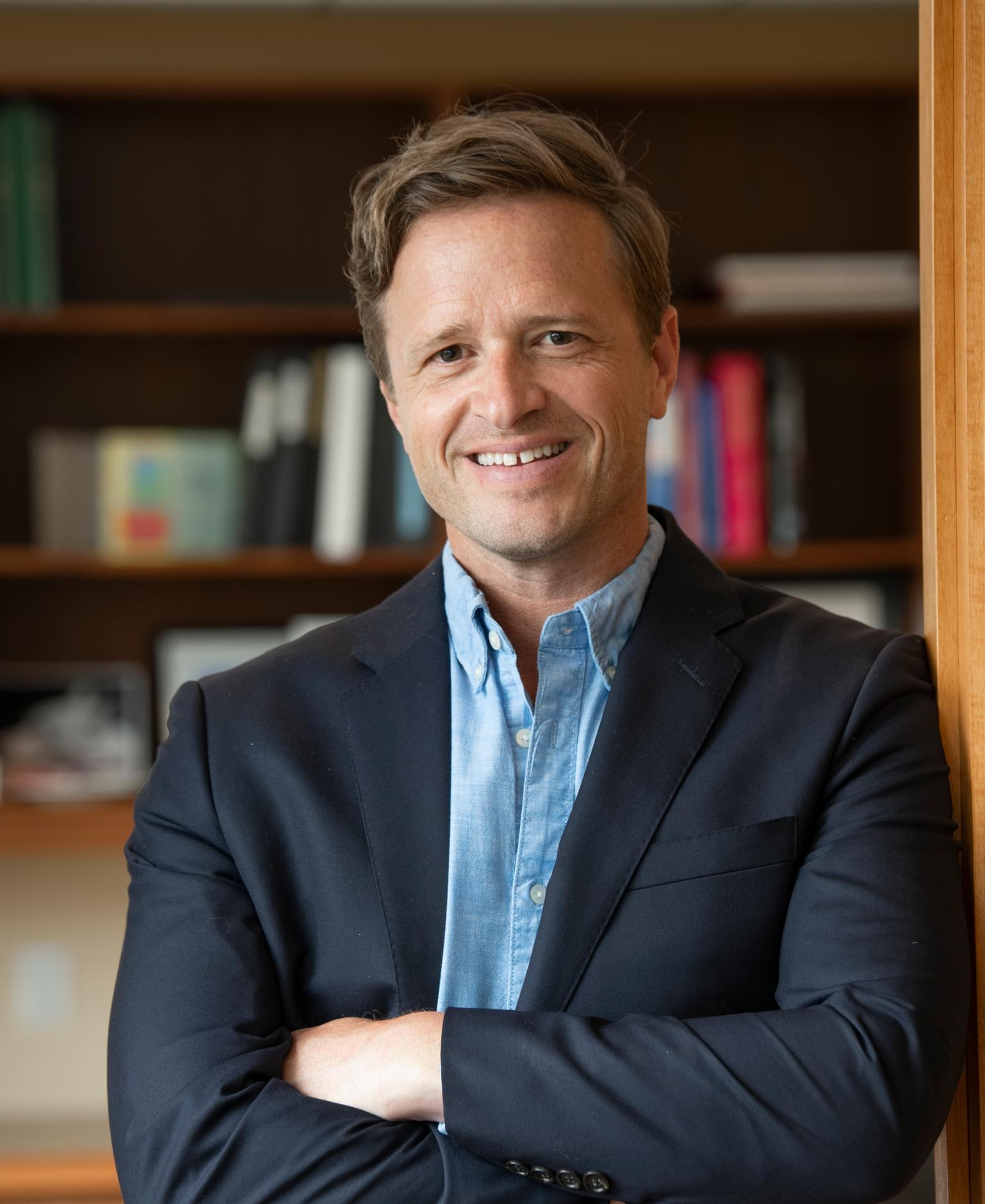
Matt Gibson, Ph.D.
Gibson’s team, led by former Stowers Postdoctoral Researcher Subu Ramanathan, Ph.D., combined live imaging of mouse intestinal organoids, miniature lab-grown models of gut tissue, with a computational model. In most tissues, the basal surface is hidden against neighboring cells or supporting material. In intestinal organoids, that surface faces outward, giving the researchers an unusually clear view of cell behavior that is normally difficult to observe.
“At the time, the lab had never worked with mice, let alone organoids,” Ramanathan said. “Matt gave me the freedom to build something outside the lab’s usual wheelhouse. That generosity is a big part of why this worked.”
Across 33 filmed cell divisions, the team observed the same overall sequence of cell rearrangements predicted by the model. Together, the approaches showed that the dramatic shape changes cells undergo during mitosis, the process by which one cell divides into two, actively reorganize neighboring cells. This revealed a fundamental mechanism that helps growing tissues maintain their structure.
Subu Ramanathan, Ph.D. Credit: University of Nebraska Medicine
“When I first imaged the basal surface in organoids, what I saw was striking: rosettes forming and neighboring cells swapping places,” said Ramanathan, who is now an Assistant Professor at the University of Nebraska Medical Center. “But the real insight came when we realized the story wasn’t that simple.”
In humans, up to 90% of cancer diagnoses are carcinomas, cancers that begin in epithelial cell layers. Understanding how these tissues normally stay organized may help researchers better understand what changes when abnormal cell growth disrupts that organization in disease.
“We now have a clearer view of how healthy epithelial tissues keep their structure as they grow,” Gibson said. “Once you understand those rules, you can start asking what changes when tissues lose that organization in disease.”
A dividing epithelial cell progresses through mitosis in a mouse intestinal organoid. The images show how the cell changes shape and position as it rounds up, divides, and produces two daughter cells within a tightly packed tissue.
How a dividing cell makes room
The findings reveal that rather than simply adding a new cell, division triggers a coordinated series of movements that changes how neighboring cells are packed. As a cell prepares to divide, it withdraws from the tissue’s basal surface, the side facing the underlying support layer. That allows neighboring cells to close in and briefly form a flower-like rosette.
“The division process creates a problem, a hole in a packed sheet, and then solves it through the geometry of the returning daughter cells,” Ramanathan said. “The shape change isn’t a side effect. It does real work.”
As the daughter cells reinsert, they trigger another round of rearrangement. Together, the two daughter cells were about 30% less circular than the original mother cell, creating a more elongated footprint that helped direct how neighboring cells rearranged, typically perpendicular to the line where the two daughter cells met.
As an epithelial cell prepares to divide, it withdraws from the basal surface, allowing neighboring cells to close in and briefly form a rosette. As the daughter cells return, they help drive another round of rearrangement, changing which cells are in contact with one another.
When researchers used a drug to arrest dividing cells before the daughter cells could return to the basal surface, neighbor exchange dropped by 93% even though rosettes still formed, showing that withdrawal and reinsertion drive distinct parts of the remodeling process.
“The neighbor exchanges weren’t happening at the same time as the rosettes, which told us our tidy story was incomplete,” Ramanathan said. “We designed an experiment to try to destroy our hypothesis, and it did. That’s what revealed that withdrawal and reinsertion drive distinct parts of the process.”
Cell division as an architect of tissue
"We tend to think of cell division as simply making two cells from one,” Ramanathan said. “This work shows that division also physically reshapes the tissue around it. Division isn’t just addition; it’s a building process.”
The finding expands how proliferation can be viewed at the tissue level. Cell division is usually described as a way to increase cell number. The new work shows that mitosis can also act as a morphogenetic process, meaning it can directly contribute to tissue form and organization.
“This study adds to a growing body of work from our lab and others which shows that epithelial proliferation is exquisitely choreographed,” Gibson said. “A dividing cell does not just become two cells. Instead, mitotic movements create geometric changes that alter the organization of the tissue around it.”
The findings also raise new questions. The researchers now want to understand how the physical properties of the surrounding tissue work together with cell geometry during division, including the role of the basement membrane and whether the same rules apply across other growing and regenerating tissues.
“Now I want to understand how the physical properties of the surrounding tissue, including the basement membrane, work together with cell geometry, and whether these rules hold across other growing and regenerating tissues,” Ramanathan said.
Additional authors include Tanmoy Sarkar, Matej Krajnc, Rosemary Mwithiga, Azize Cerci, and Chongbei Zhao.
About the Stowers Institute for Medical Research
Founded in 1994 through the generosity of Jim Stowers, founder of American Century Investments, and his wife, Virginia, the Stowers Institute for Medical Research is a nonprofit biomedical research organization with a focus on foundational research. Its mission is to expand our understanding of the secrets of life and improve life’s quality through innovative approaches to the causes, treatment, and prevention of diseases. The Institute consists of more than 20 independent research programs. Of the approximately 500 members, over 370 are scientific staff that include principal investigators, fellows, technology center directors, postdoctoral scientists, graduate students, and technical support staff. Learn more about the Institute at www.stowers.org and about its graduate program at www.stowers.org/gradschool.
Media Contact Joe Chiodo, Director of Communications Stowers Institute for Medical Research 724-462-8529 jchiodo@stowers.org