The detachment of neural crest cells occurs via a process called
epithelial to mesenchymal transition—where static cells change their
shape and acquire migratory properties—the subject of a recent review paper
from the Trainor Lab. Once situated in the facial region, neural crest
cells differentiate, or decide their cell fate, ultimately generating
nearly all the of the craniofacial cartilage, bone and connective
tissues.
The rapid proliferation and growth of neural crest cells in concert
with epithelial to mesenchymal transition requires a disproportionate
quantity of ribosomes and hence proteins compared to surrounding
non-neural crest cells and tissues. One of the key steps of ribosome
biogenesis—RNA Polymerase I-mediated rRNA transcription—is a
rate-limiting step in the formation of ribosomes, and is highly active
within neural crest cells.
“Large quantities of protein translation are needed for neural crest cell proliferation and survival,” said Dash.
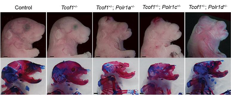
By turning off genes responsible for rRNA transcription and ribosome
formation in mice, the researchers uncovered a cellular imbalance
between rRNA transcription and ribosomal proteins that greatly impacted
neural crest cells.
This imbalance promotes neural crest cell death via increased
concentrations of the tumor suppressor protein, p53, which causes the
embryo to develop malformations of the head and face.
In normal neural crest cells, p53 levels are tightly controlled by
the protein, Mdm2. However, when rRNA transcription is reduced, an
imbalance arises where ribosomal proteins that normally form part of the
ribosome, become free and instead bind to Mdm2 preventing its control
of p53. This leads to increased levels of p53 that cause the newly
formed neural crest cells to die.
“We have known for many years that problems in craniofacial
development are often the result of neural crest cells dying before they
can form the cartilage and bone structures of the head and face,” said
Falcon.
“Our previous studies have shown that inhibiting p53 in
Treacher-Collins Syndrome animal models can stop neural crest cells from
dying and thus prevent craniofacial anomalies from occurring, but this
made the animals much more susceptible to cancer,” said Watt.
“The significance of this study is that at the genetic, cellular, and
biochemical level, we can now connect rRNA transcription and the
ribosome biogenesis pathway in neural crest cells, to proper
craniofacial development and to the pathogenesis of birth defects,” said
Trainor. “We understand how each step in the pathway links to
p53-dependent apoptosis.”
Coauthors include Ruonan Zhao, Daisuke Sakai PhD, Emma L. Moore,
Sharien Fitriasari, Melissa Childers, Mihaela E. Sardia, Selene Swanson,
Dai Tsuchiya, Jay Unruh, PhD, George Bugarinovic, Lin Li, Rita Shiang,
PhD, Annita Achilleos, PhD, Jill Dixon, PhD, and Michael Dixon, PhD.
Studying multiple research organisms is key
The study of rare diseases is challenging but deserves no less
attention than common diseases. However, a universal mechanism to
explain rare birth defects simply doesn’t exist and developing
preventative therapies therefore requires a thorough understanding of
the cellular and genetic mechanisms responsible for each individual
disorder, which is derived from our knowledge of normal craniofacial
development in many different animal species.
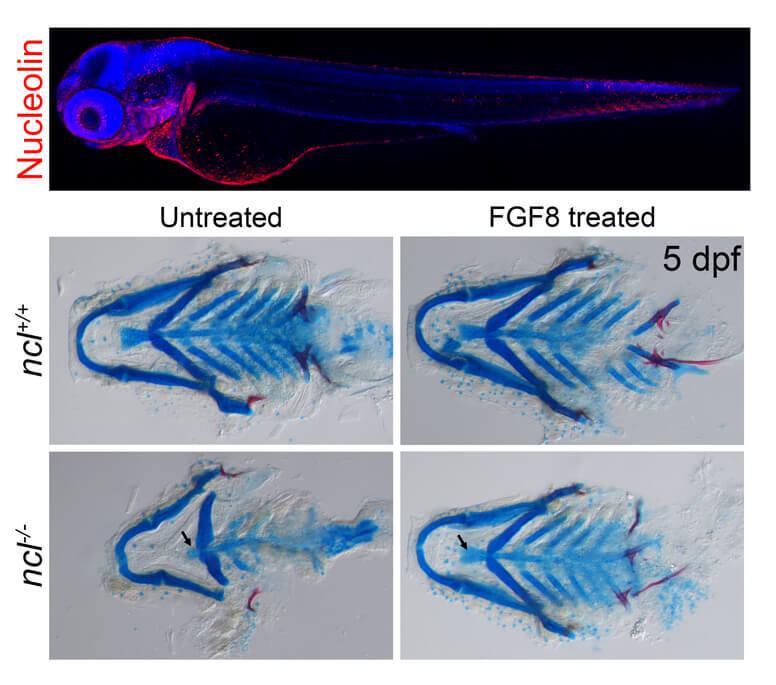
For example, in another recent study published in Development
on June 28, 2022, Dash in the Trainor Lab investigated the function of
Nucleolin in neural crest cells and craniofacial development in
zebrafish. Mutations in Nucleolin disrupt the differentiation of neural
crest cells resulting in craniofacial anomalies resembling
Treacher-Collins Syndrome. Dash discovered that Nucleolin, among its
many functions, regulates signaling pathways that are important for
neural crest cell differentiation into bone and cartilage.
“By identifying the biochemical pathway that was involved, we were
able to bypass the problem by providing the zebrafish embryos with the
specific proteins they needed for proper differentiation,” said Dash.
Using research organisms such as mice and zebrafish to understand the
basis for birth defect disorders can lead to discoveries on how to
prevent them. “If it’s successful in animal models, it may give people
hope that something can be done in the future,” said Trainor.
This work was funded by the Kirschstein-NRSA F31 predoctoral
fellowship (awards DE027860, DE023017) from the National Institute for
Dental and Craniofacial Research of the National Institutes of Health,
K99 Pathway to independence awards (DE030971 and DE030972), the American
Association for Anatomy Post-Doctoral Fellowship, and from institution
support from the Stowers Institute for Medical Research. The content is
solely the responsibility of the authors and does not necessarily
represent the official views of the NIH.