News


22 July 2026
Coral Keepers
Inside our coral spawning success. In the heart of the midwest.
Read Article
News
For Postdoctoral Research Associate Joseph Varberg, researching proteins in yeast is more fascinating through the lens of a microscope.
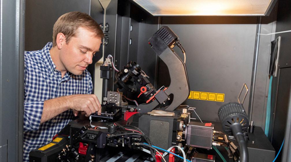

For Postdoctoral Research Associate Joseph Varberg, researching proteins in yeast is more fascinating through the lens of a microscope, and he’s noticed it’s true for others, too.
“There’s just something about being able to see it all that makes it that much more interesting,” he said. “If I’m talking about my project with my friends or family, they may not follow, but I can show them images, and it clicks. Oftentimes, when they see the images, they ask some of the same questions that we, the scientists, are interested in.”
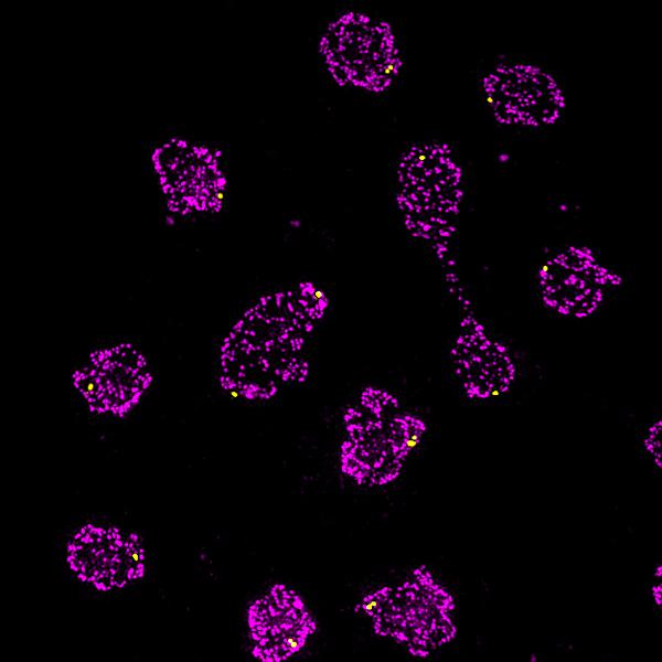

Fission yeast Schizosaccharomyces pombe.
Image courtesy of Joseph Varberg.
Since starting his postdoc at the Stowers Institute in 2017, Varberg has used imaging techniques as a focus for his research on proteins in the fission yeast Schizosaccharomyces pombe. Yeast is a valuable tool for studying fundamental cellular processes shared by many organisms, including humans. And fission yeast cells, which grow exclusively through their tips and divide across the middle to create two daughter cells of equal size, are particularly useful for studying mechanisms of cell growth and division. Understanding how the cells accomplish these biological feats normally is critical for determining what happens when they malfunction, which can lead to a wide spectrum of human diseases, including cancer and birth defects.
Each cell’s genetic material is surrounded by a protective barrier called the nuclear envelope, and this is where Varberg’s research lies. He studies a specialized set of proteins in the nuclear envelope that help maintain the shape and function of the nucleus—thus protecting the instructions for life contained within—and also influence chromatin organization and gene expression.
In 2019, Varberg was awarded an F32 postdoctoral fellowship from the National Institutes of Health, and he set out to screen a library of S. pombe membrane proteins and develop imaging and analysis methods to characterize the organization of the yeast nucleus. As he’s specifically interested in studying proteins at the inner membrane of the nuclear envelope—and previous tools for looking at protein-protein interactions don’t work with membrane proteins—he used a special approach for this project called the “membrane yeast two-hybrid” system, or MYTH, to perform high-throughput screening and identify network interactions for three nuclear envelope proteins of diverse structure and function. “By building a list of proteins and complexes present at the inner nuclear membrane, we hope to expand our current understanding of how these proteins interface with the genome as well as how they perform essential functions of the nucleus,” said Varberg.
The results of his research were published in December 2020 in the journal G3, providing a tool for studying membrane protein interactions for the S. pombe community. This will support further studies of nuclear envelope proteins in fission yeast as well as comparative studies in other research systems.
Varberg began his postdoc career in the Jaspersen Lab and then moved to the Gerton Lab, which has a similar focus on the biology of the nucleus. He’s developing new ideas with the team but imaging still features heavily in his research, and he continues to work closely with the Microscopy Center team.
“That’s been one of my major focuses—utilizing various microscopy techniques and developing tools to analyze the resulting data,” Varberg said, adding that “with the technology, resources, and training available from the Microscopy Center, you can quickly move from having a hypothesis to having data in hand.”
Varberg’s fascination with biology developed gradually. In high school Varberg focused on literature, history, and music. But during a chemistry class in his sophomore year of college, he realized he had a knack for the subject. He switched his major to chemistry and began working on a collaborative research project conducting computational modeling of new polymer materials for solar devices.
During a summer internship at the University of Kansas, he delved deeper into experimental work, using ultrafast spectroscopy to study the properties of compounds being developed for use in next-generation ultra-high-density data storage devices.
While finishing his bachelor’s degree, he began working as a technician in a lab at the Stowers Institute, where he pursued molecular and cell biology. He enjoyed the pace of laboratory bench work, and his doctoral plans shifted from chemistry to biology. At the Indiana University School of Medicine, he was co-mentored by two investigators working on the parasite Toxoplasma gondii.
During his doctoral research Varberg focused on understanding the machinery used by the parasite to sense and respond to cellular stresses, and how misregulation could make the parasites die, with the aim of identifying potential new drug targets for treatment of toxoplasmosis and other pathogenic diseases.
Going forward, Varberg will continue to hone his coding skills, and he’s also doing more live-cell imaging. In the Gerton Lab, he's testing the functional significance of the interactions for proteins present in the inner nuclear membrane. Historically, even identifying proteins on the inside of the nuclear envelope, versus what’s on the outside, has been a challenge, he said.
“Now that we’ve identified these proteins, we can begin to study how they impact nuclear organization and function using our imaging approaches,” Varberg said. “The next step is to continue to develop those imaging and analysis tools to be able to test our hypotheses in a quantitative way.”
News
22 July 2026
Inside our coral spawning success. In the heart of the midwest.
Read Article
News
22 July 2026
Dive into the biological basics of symbiosis, the close partnerships that allow different organisms to thrive together, and take a closer look at how a Stowers scientist explores the molecular conversations that make these relationships possible.
Read Article
News


17 July 2026
Evan Morrison, Ph.D., a Stowers Institute postdoctoral researcher and a 2021 recipient of the Howard Hughes Medical Institute Gilliam Fellowship, followed an unconventional path into biological research, and continues his HHMI fellowship at the Stowers Institute researching how cells identify defective ribosomes.
Read Article


