News


22 July 2026
Coral Keepers
Inside our coral spawning success. In the heart of the midwest.
Read Article
News
Modeling the makeup and mechanism of cnidarian stingers
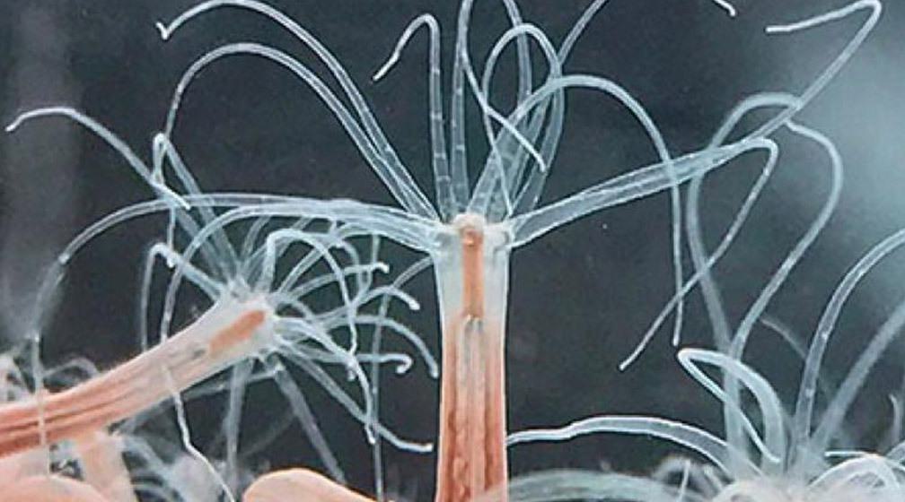

The stinging starlet sea anemone, Nematostella vectensis.
By Rachel Scanza, PhD
Death and taxes. The two inevitabilities of life. Or are they? Obviously, the starlet sea anemone doesn’t pay taxes, at least not in the traditional sense. However, this animal just happens to be immortal. The absence of senescence—the unavoidable decline in cellular and organ function with time—along with regenerative capabilities has established the cnidarian sea anemone as a valuable research organism for over a century. In addition, the stinging mechanism by which a sea anemone detects, releases, penetrates, and poisons its victim occurs in just a few thousandths of a second, making it one of the fastest processes occurring in nature.
Cnidarians are a group of water-dwelling sea creatures, ranging from corals to jellyfish. Wildly diverse in morphology, and with an evolutionary history dating back between 500-700 million years, these animals share similar stinging structures typically used for predation and protection. The starlet sea anemone, Nematostella vectensis, possesses a complex organelle called a nematocyst manufactured from a specialized cell, a nematocyte.
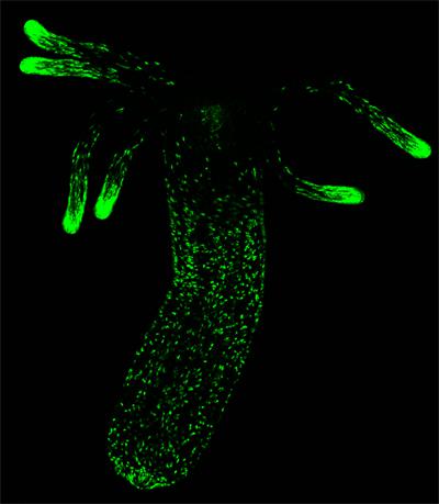

Fluorescent expression of nematocytes, the specialized cell that creates nematocysts. Notice that nematocyte concentration is highly expressed in the tentacle tips.
Cnidarian nematocysts have been the subject of scientific study for over 100 years and while the general structure is understood, its sophistication and the speed and complexity of the stinging mechanism remained elusive. The exponential increase in technological advances now makes it possible to precisely underpin nematocyst form and function. New research from the Stowers Institute for Medical Research has resolved a complete model for the architecture and operation of the cnidarian stinging organelle at unprecedented spatial and temporal resolution. The findings can aid scientists studying different types of cnidarians and may have applications for designing medical delivery devices.
The nematocyst is comprised of a capsule containing a stinging thread and is sealed by three flaps. Two sub-structures define the thread: a rigid shaft and a long tubule dotted with barbs. Armed with an arsenal of technologies and techniques, Predoctoral Researcher Ahmet Karabulut from the lab of Matt Gibson, PhD, in collaboration with Melainia McClain, Boris Rubinstein, PhD, and Sean McKinney, PhD, from the Stowers Institute Technology Centers revealed in detail nematocyst structure and characterized the complex firing sequence into three unique time phases in a recent study published in Nature Communications on June 17, 2022.
“Although stinger biology has been studied for a long time, modern techniques have now allowed us to uncover the intricate details about the structure and process that were not previously known,” said Karabulut.
When triggered, a nematocyst responds with both rapidity and precision. The energy required for the initial thread discharge arises from osmotic pressure, the unequal distribution of charged molecules between the inside of the capsule and its surroundings, and elastic energy generated when the capsule wall stretches. The speed at which the thread is ejected from the capsule is acquired by a sudden influx of ion-neutralizing water that results in the capsule doubling in volume, converting osmotic pressure and elastic energy to kinetic energy sufficient to puncture its target. Subsequent shaft and tubule elongation additionally convert elastic energy stored in both the thread wall and in connector regions into kinetic energy.
To visualize the resting structure of the capsule and thread and how it evolves during firing, Karabulut and his coauthors introduced three types of fluorescent tags. The thread’s shaft is composed of three filaments tightly coiled in a helical conformation. Fluorophore expression allowed visualization of both the thread as well as a neuron-like network connecting individual threads; these synaptic networks may act to coordinate nematocyte activity.
A three-dimensional reconstruction of nematocyst architecture was created to study the structure and functionality of the shaft and tubule. Along with fluorescent tagging, the researchers sampled sections of a resting nematocyst and used a scanning electron microscope to magnify each section. Filaments are compactly coiled and oriented parallel to the opening of the capsule in contrast to the propeller-shaped cross sections of the tubule. In addition, two connector regions could be identified, one region connecting the flaps covering the top of the capsule to the shaft and a similar region at the base of the shaft which connects to the twisted tubule.
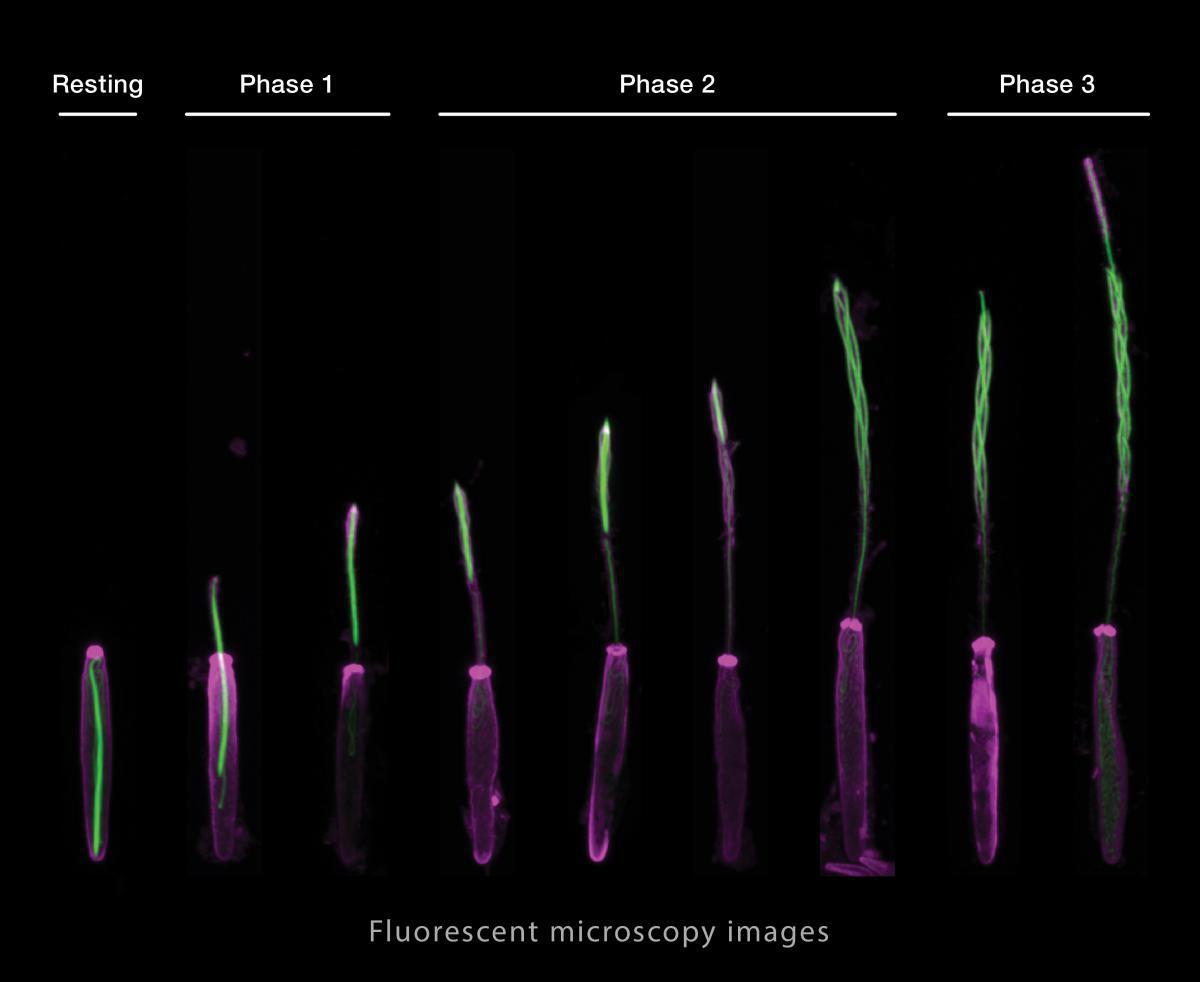

Fluorescent microscopy images illustrate the three phases of thread eversion during nematocyst firing.
When a nematocyst discharges, the stinging thread emerges from the capsule and is propelled forward and elongated by a mechanism called eversion, where the thread substructures turn inside out. Recording the fluorescent discharge events at high speeds, Karabulut was able to define that nematocyst firing occurred in three unique phases. Phase 1, 2 and 3 are characterized as shaft discharge, shaft eversion, and tubule eversion, respectively.
“The new techniques described in our paper are a foundation for future studies that couldn’t have possibly been done in the past,” said Karabulut.
During phase 1, the shaft is ejected from the capsule as a dense missile-like structure in a highly coiled configuration. The shaft is propelled forward until the shaft-capsule connector is stretched to its maximum length. This marks the beginning of phase 2 when the three filaments of the shaft start to uncoil and turn inside out at the top of the shaft, allowing the bottom of the shaft to move forward within the uncoiling filaments. The tubule that is connected to the base of the shaft is also pulled forward within the cavity created by eversion of the shaft.
When the tubule emerges from the everted shaft (Phase 3) it begins its own process of eversion where elastic energy is converted to kinetic energy as the tubule wall unfolds and untwists, forming a hollow cylinder. The tip of the elongating tubule is dotted with barbs that not only deliver venom to its target but also provide structural support during elongation.
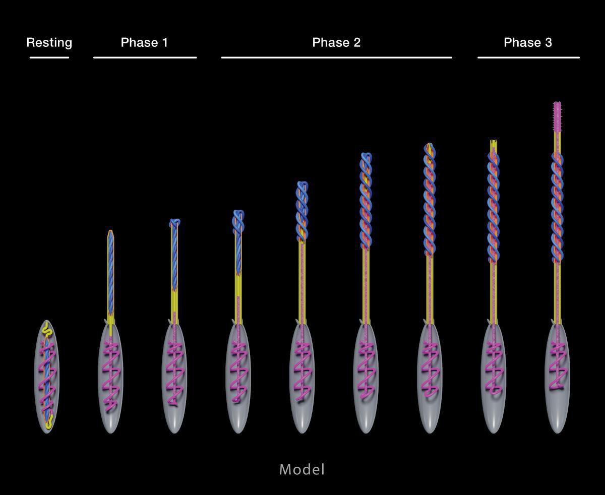
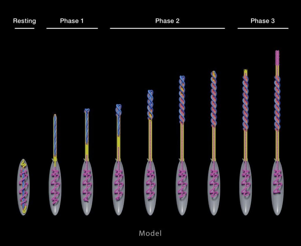
Model shows precise mechanism of eversion during each nematocyst firing phase.
“There are such orchestrated layers of a beautiful structure like this with very complex transformations,” said Karabulut. “If we can understand these, in the future we can make devices like this that could deliver targeted medicines in humans.”
For the first time, the structure and mechanism of thread firing and eversion has been characterized with extraordinary detail in a cnidarian stinging organelle, enabling the construction of a precise nematocyst model. The stinging operation occurs in three stages where the complex transformation and elongation of the thread substructures is coupled to various forms of stored energy which are converted to kinetic energy required for the process. The model is a valuable resource for scientists studying different cnidarian species and may provide a template for constructing micro-medical delivery devices.
“This new knowledge is the product of the exploratory environment of the Stowers Institute,” said Gibson. “Curiosity-driven fundamental research can often lead to really exciting new ideas or applications.”
Coauthors include Melainia McClain, Boris Rubinstein, PhD, Keith Z. Sabin, PhD, and Sean McKinney, PhD.
Funding for the study was provided by institutional support from the Stowers Institute for Medical Research.
News
22 July 2026
Inside our coral spawning success. In the heart of the midwest.
Read Article
News
22 July 2026
Dive into the biological basics of symbiosis, the close partnerships that allow different organisms to thrive together, and take a closer look at how a Stowers scientist explores the molecular conversations that make these relationships possible.
Read Article
News


17 July 2026
Evan Morrison, Ph.D., a Stowers Institute postdoctoral researcher and a 2021 recipient of the Howard Hughes Medical Institute Gilliam Fellowship, followed an unconventional path into biological research, and continues his HHMI fellowship at the Stowers Institute researching how cells identify defective ribosomes.
Read Article


