News


22 July 2026
Coral Keepers
Inside our coral spawning success. In the heart of the midwest.
Read Article
News
Q&A with Tom Kleist, Ph.D, a member of the Institute’s Microscopy Technology Center
What do you think people would find most interesting about your job and the technology you use?
Biology is oftentimes abstract. It can be challenging for a general audience to understand plots summarizing large-scale ‘omics projects, graphs showing mass spectrometry results, or tree diagrams showing inferred relationships among genes or organisms. Microscopy is totally different and refreshingly tangible. We use many different advanced techniques to label and visualize molecules and cells; but, in essence, the results are images of physical structures that are simply too small to see by eye. The tangible nature of microscopic images makes it easier for general audiences to appreciate these and therefore to attract their interest.
What exactly is microscopy?
Microscopy is the technical discipline of using microscopes to image very small structures that we simply cannot see by eye. There are many types of microscopy, which can be broadly categorized as either light microscopy or electron microscopy. In light microscopy, beams of light are used to illuminate and magnify the sample. In electron microscopy, beams of electrons are used instead. The new microscope in the Özel Lab is an advanced type of light microscope that is specially designed for high-resolution imaging of fluorescent molecules in biological samples.
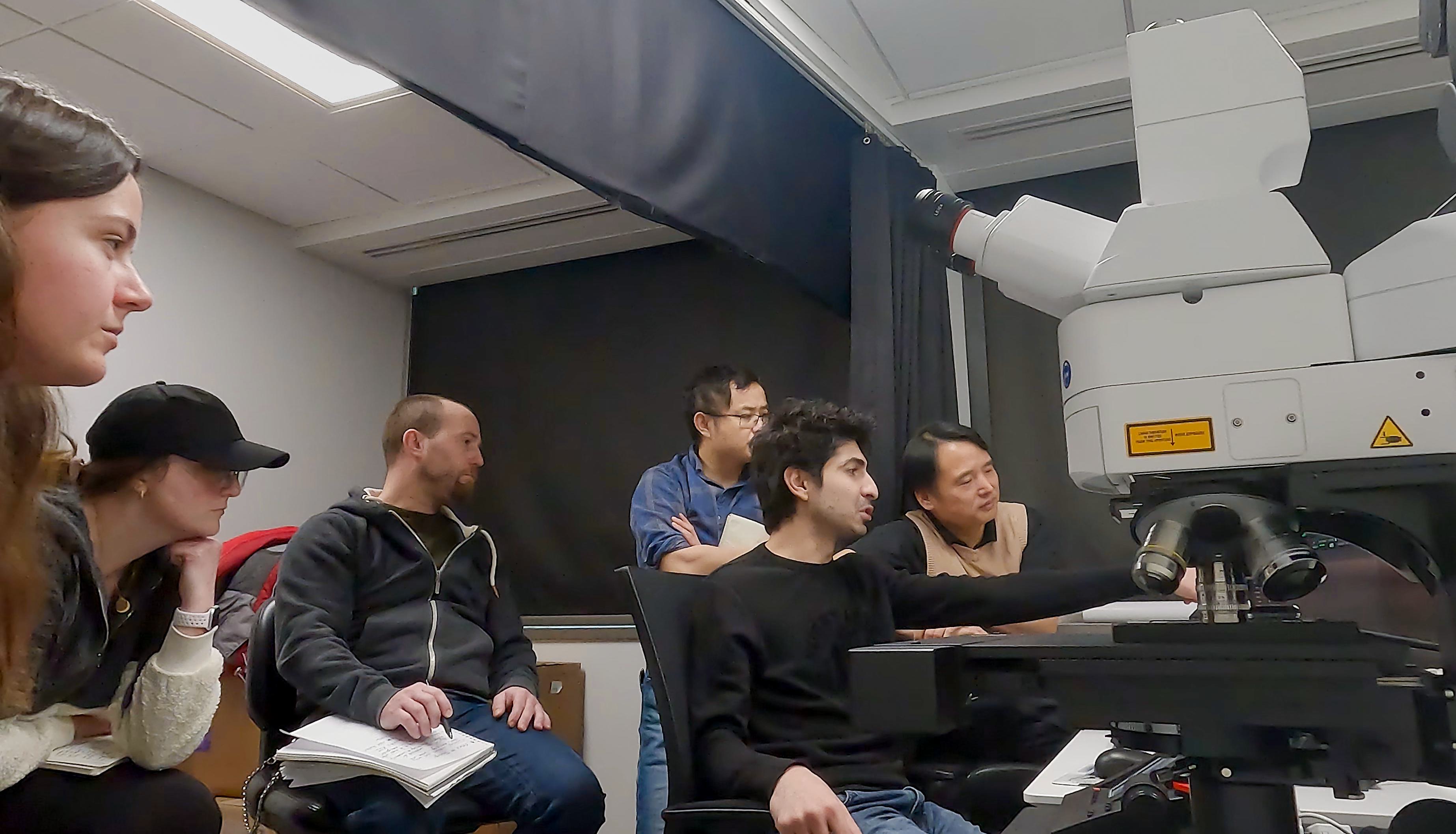
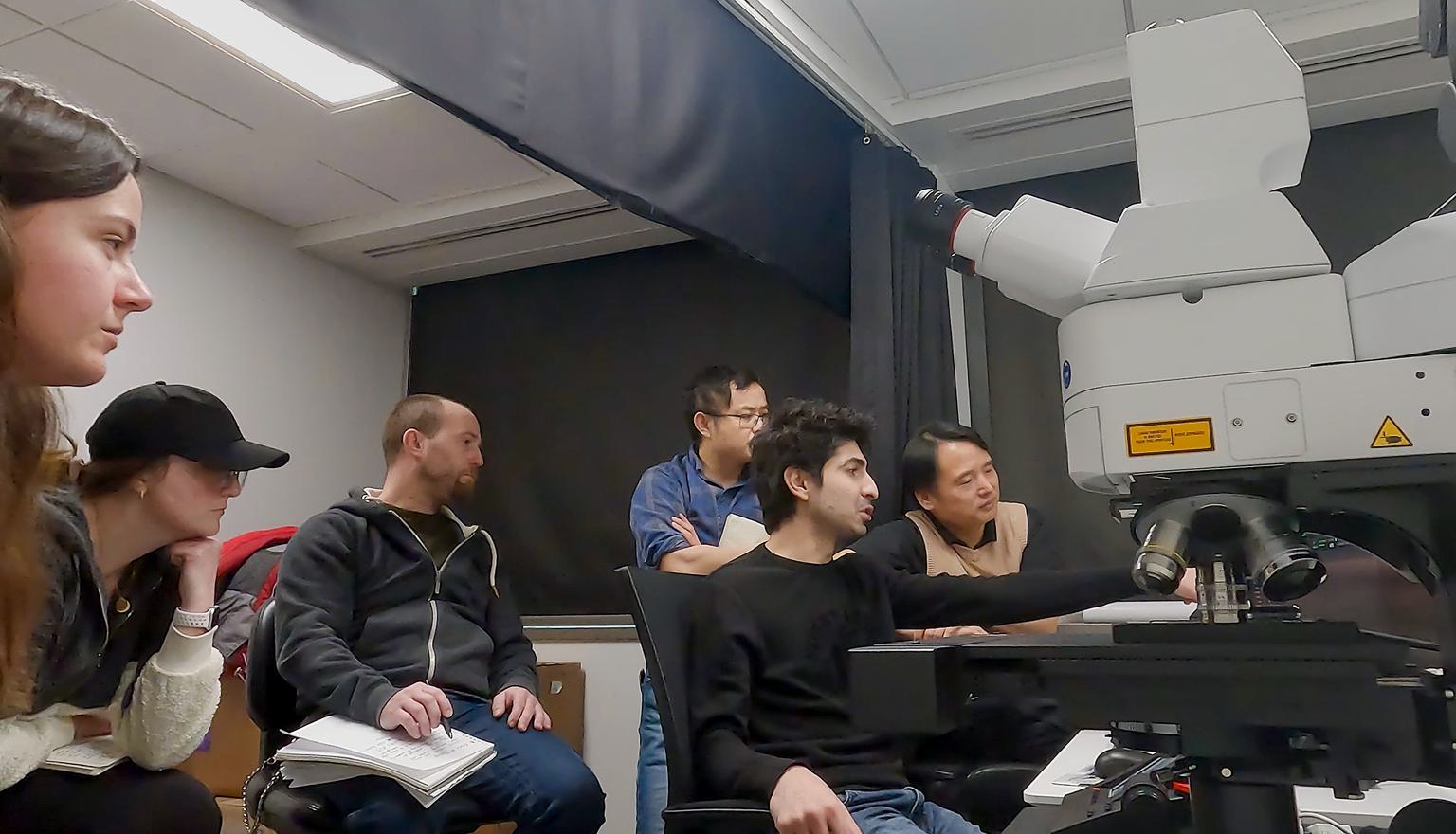
Members of the Özel Lab training on the new microscope
What is this new microscope, and what does it do differently than other microscopes we have at the Institute?
The Özel Lab’s Leica Stellaris 8 is an upright confocal fluorescence microscope. It contains a novel tunable white light laser that allows users to specify any illumination wavelength from 440 – 790 nanometers, instead of the usual four or five fixed wavelengths available on most microscopes. The white light laser covers most of the visible spectrum and into the infrared spectrum, so researchers can tune the laser for optimal sample viewing. The microscope has new, extremely sensitive detectors that can pick up even faint amounts of light, which is important because intense illumination tends to degrade samples. The microscope includes customized software that can deconvolve images for improved spatial resolution – essentially it has an algorithm that corrects for distortions caused by the refractive or “bendy” nature of light as it passes through different media. Lastly, the upright format of the microscope allows users to perform imaging of live fruit flies – the Özel Lab’s major research organism – in their natural standing orientation. Otherwise, the flies would be upside down!
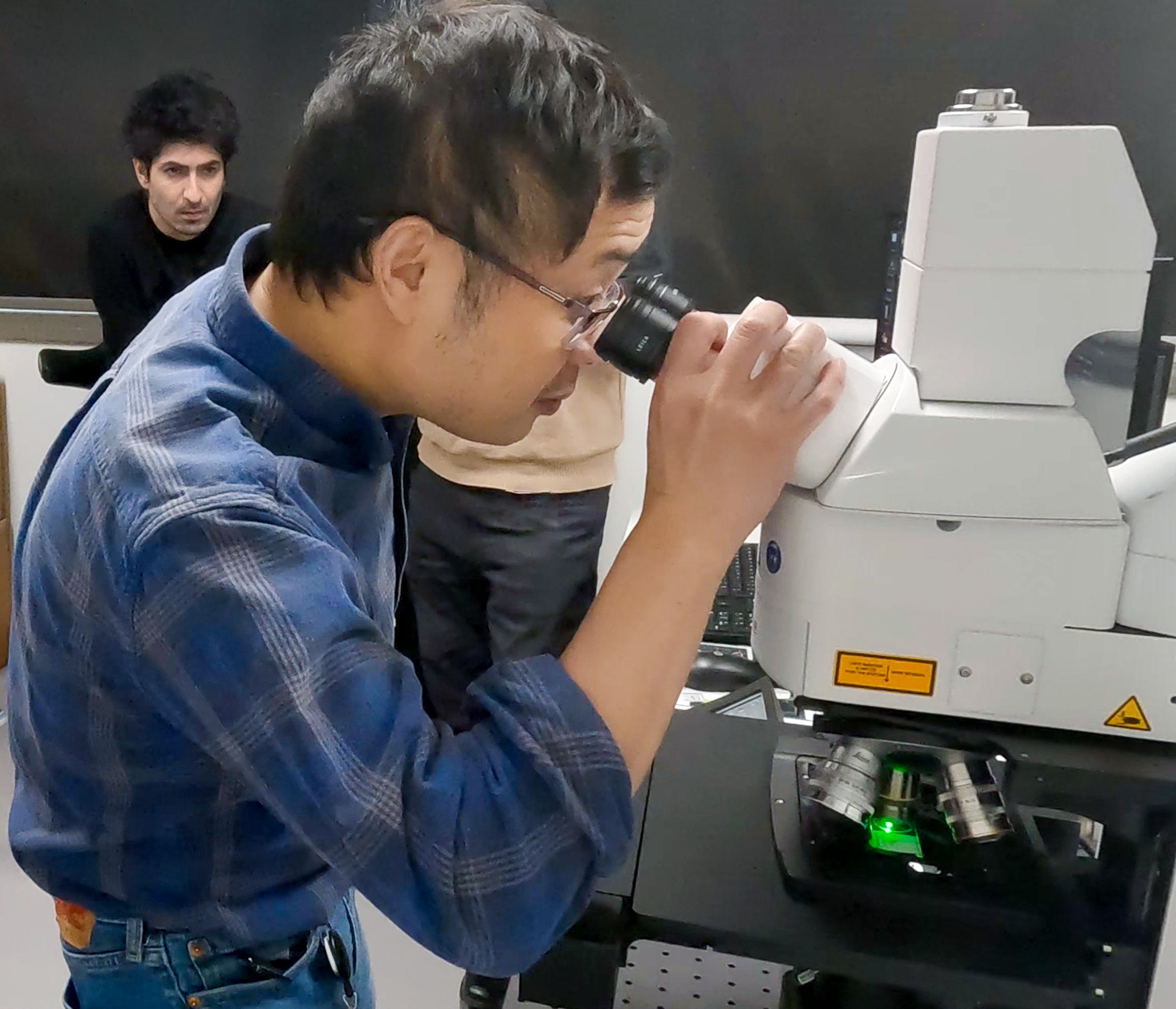

Institute member Zulin Yu, Ph.D., the Leica Stellaris 8 microscope
What is the benefit of having this microscope at Stowers?
This microscope is the perfect tool for work in the Özel Lab. It will enable them to image entire fly brains with subcellular resolution. Looking forward, this scope will allow them to image neural activity in brains of living flies – a demanding type of experiment that requires highly advanced technology.
What are some of the most exciting discoveries achieved with this technology?
Confocal fluorescence microscopy has truly transformed biology. There are too many exciting discoveries to list. During his postdoctoral training, Neşet published multiple papers in the journals Nature and Science that relied on similar, slightly older microscope technology. Neşet’s research has been groundbreaking in revealing how neurons are developmentally specified in the fly brain, which helps us to better understand principles that underpin neurological development of humans too.
News
22 July 2026
Inside our coral spawning success. In the heart of the midwest.
Read Article
News
22 July 2026
Dive into the biological basics of symbiosis, the close partnerships that allow different organisms to thrive together, and take a closer look at how a Stowers scientist explores the molecular conversations that make these relationships possible.
Read Article
News


17 July 2026
Evan Morrison, Ph.D., a Stowers Institute postdoctoral researcher and a 2021 recipient of the Howard Hughes Medical Institute Gilliam Fellowship, followed an unconventional path into biological research, and continues his HHMI fellowship at the Stowers Institute researching how cells identify defective ribosomes.
Read Article


