News


22 July 2026
Coral Keepers
Inside our coral spawning success. In the heart of the midwest.
Read Article
News
Steph Nowotarski is a scientist in the Stowers Institute’s Microscopy Technology Center.
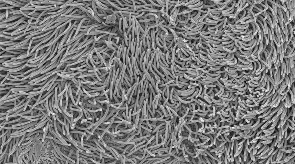

Cilia on planarian pharynx. Image courtesy of Melainia McClain
What do you think people would find most interesting about your job and the technology you use?
We can’t understand how biology works just by studying it when it goes wrong. We need to understand how things work normally before focusing on fixing. For example, I’d have to know how a carburetor works to fix one in my car and first I’d need to know what each part looks like and where they fit. Microscopy is a powerful tool to understand and visualize where important structures and players in biology are and electron microscopy lets us see the tiniest of details and how they fit into the bigger picture of biology.
What exactly is microscopy?
Just like how a magnifying glass makes samples bigger using light, the electron microscope uses magnetic fields (instead of glass lenses) to focus electrons (instead of light). Because electrons are smaller than light, we can see structures that are smaller than what we can see with any light microscope, from a magnifying glass to a top-of-the-line light microscope.

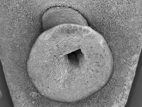
Planarian pharynx. Image courtesy of S. Nowotarski.
What does the Merlin SEM with 3View do?
The Zeiss Merlin SEM at Stowers is like a very, very expensive pocketknife…that’s way too big to fit in your pocket! It can do three different things. First, it can take very detailed images of the outside of a specimen, giving fine details about the surface of samples. This mode looks like super, super, super macro photography.
The Zeiss Merlin can also look at the inside of samples. To do this we cut a specimen into super thin slices with a diamond knife after it has been embedded in plastic. How thin? Like so thin that it would take 800 slices to capture the width of the average human hair!
The power of this machine in these modes isn’t solely based on how you can see super tiny things, but that you can take large, sweeping fields of view and then zoom in to high resolution wherever you want, like Google Maps . This lets us and our researchers pinpoint regions of interest and allows us to align our cell-level biology into the context of a whole animal.
The last thing it can do with an addition of the 3View module is what technically makes it a knife (but still too big for a pocket!). It can take an image of a sample, cut off a thin section of it with a diamond knife that is in the microscope, and then image the surface the knife revealed. It does this repeatedly, sometimes running for multiple weeks 24/7, and at the end, we get a 3D volume of high-resolution data.
From outside to inside, and from 2D to 3D, the Zeiss Merlin with 3View brings tiny biology into perspective!
3D structure of neuromast-associated ionocyte (Nm ionocytes).
Image courtesy of Melainia McClain from Peloggia et. al, 2021)
What is the benefit of having this technology at Stowers?
Light microscopy and electron microscopy are synergistic technologies. Each fills the other’s weaknesses. Light microscopy is limited by physics and markers. We can only resolve things that are a certain distance apart. It used to be the limit was ~250 microns; now, with super-resolution we can get down an order of magnitude to ~40 nm (or 0.04 microns). We also must be able to use chemicals to stain tissues, or fluorescent proteins to mark proteins to make them stand out and provide enough contrast. Electron microscopy can get down another order of magnitude and resolve structures that are only angstroms apart (~0.0001 micron). The Zeiss merlin, for what we generally do here at Stowers looks at structures on the order of 5-50 nm. While it too relies on staining to create contrast, unlike staining techniques in light microscopy, the staining allows us to see all the surrounding structures. This allows our researchers to look at and understand biology on the smallest of scales but also allows us to zoom out and put those tiny details into perspective.
What are some of the most exciting discoveries achieved with this technology?
Scanning electron microscopy looking at the outside of specimens allows scientists to fully understand the normal state of a tissue or animal and then we use it again to see what happens when we remove the function of a gene, or when we turn a gene on higher, or in the wrong place.
Looking at a set of thin sliced sections on this machine allows us to marry the best of light microscopy and electron microscopy by combining them with correlative light and electron microscopy (CLEM). We can see a cell’s molecular identity (light microscopy) and its full structure (electron microscopy).
The technology of 3D electron microscopy that the 3View offers is relatively new and it is allowing scientists to understand biology at the smallest of scales in the context of a whole 3D tissue. Recently, a groundbreaking paper captured a whole animal at high resolution using this technology, providing an atlas for their field. Here at Stowers, we are using it to understand what stem cells and their local environments look like, as well as using it to characterize and understand cells and tissues in new research organisms like sea anemones and apple snails! This new technology is just starting to take off and promises a lot more to come!
News
22 July 2026
Inside our coral spawning success. In the heart of the midwest.
Read Article
News
22 July 2026
Dive into the biological basics of symbiosis, the close partnerships that allow different organisms to thrive together, and take a closer look at how a Stowers scientist explores the molecular conversations that make these relationships possible.
Read Article
News


17 July 2026
Evan Morrison, Ph.D., a Stowers Institute postdoctoral researcher and a 2021 recipient of the Howard Hughes Medical Institute Gilliam Fellowship, followed an unconventional path into biological research, and continues his HHMI fellowship at the Stowers Institute researching how cells identify defective ribosomes.
Read Article


