What exactly is light microscopy?
Primarily, light microscopy utilizes fluorescent dyes or proteins and
specialized light sources to visualize samples. In a fluorescence
microscope, a specific wavelength of light is used to illuminate the
sample, causing the fluorescent molecules to emit light at a different
wavelength. Detectors then capture the emitted light, which is used to
produce an image of the sample.
The most frequently used fluorescent microscopes at Stowers are laser
scanning and spinning disk confocal systems. Laser scanning confocal
systems utilize a focused beam of light to scan the sample while
pinholes block out-of-focus light, resulting in clearer images. This
type of confocal system is particularly useful for studying the 3D
structure of biological samples. Spinning disk systems are similar to
laser-scanning confocal systems but are 100 times faster. This makes
them more suitable for imaging large samples and volumes, and live
samples where speed is essential.
Technologies at our center include super-resolution microscopes that
can visualize structures even smaller than the wavelength of light.
What does the SIM microscope do?
Conventional light microscopy is limited by the diffraction of light,
which restricts the ability to resolve structures smaller than
approximately half the wavelength of light used to illuminate the sample
(around 200 nm).
To overcome this, super-resolution microscopy techniques have been
developed that use a variety of approaches to manipulate the interaction
between light and the sample. These techniques include structured
illumination microscopy (SIM), stimulated emission depletion (STED)
microscopy, and single-molecule localization microscopy (SMLM).
The SIM microscope works by using patterns of light to illuminate the
sample. An algorithm is applied to process the resulting images,
allowing the SIM microscope to create a high-resolution image of the
sample with details beyond the diffraction limit. With SIM, a resolution
of up to twice the diffraction limit of conventional light microscopy
can be achieved, making it a powerful tool for studying fine details of
cells such as microtubules and the nuclear envelope.
What is the benefit of having this technology at Stowers?
SIM offers several advantages over other super-resolution technologies.
Firstly, it is a versatile tool that can work with various conventional fluorescent proteins and dyes, making it suitable for many researchers at Stowers. Additionally, SIM can capture a large field of view simultaneously, making it faster than other super-resolution techniques like STED microscopy. SIM can also be used to image live samples, allowing researchers to study dynamic biological processes in real-time. This makes it particularly useful for investigating cellular processes such as cell division, migration, and signaling.
What are some of the most exciting discoveries made so far with this technology? Both at Stowers and elsewhere.
SIM has enabled researchers to make many exciting discoveries that
were previously impossible with traditional light microscopy since its
development two decades ago. According to a PubMed analysis, there have
been over 2,000 publications per year in recent years that use SIM,
indicating that it has become a widely used tool to answer important
questions in biological research.
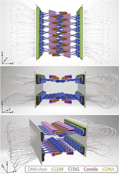
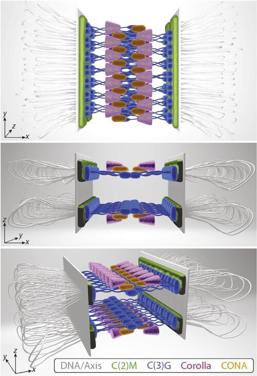
At the Stowers Institute, Investigator Scott Hawley and his
colleagues were the first to use SIM after the technology became
available in 2013. They used SIM to image the synaptonemal complex (SC),
a protein structure that facilitates the exchange of genetic material
between homologous chromosomes during meiosis. The high-resolution
images obtained using SIM allowed the researchers to better understand
the structure and function of the SC. More recently, the lab presented an
updated SC model based on new data from SIM, revealing two layers of SC
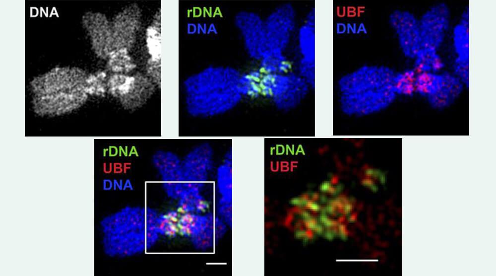
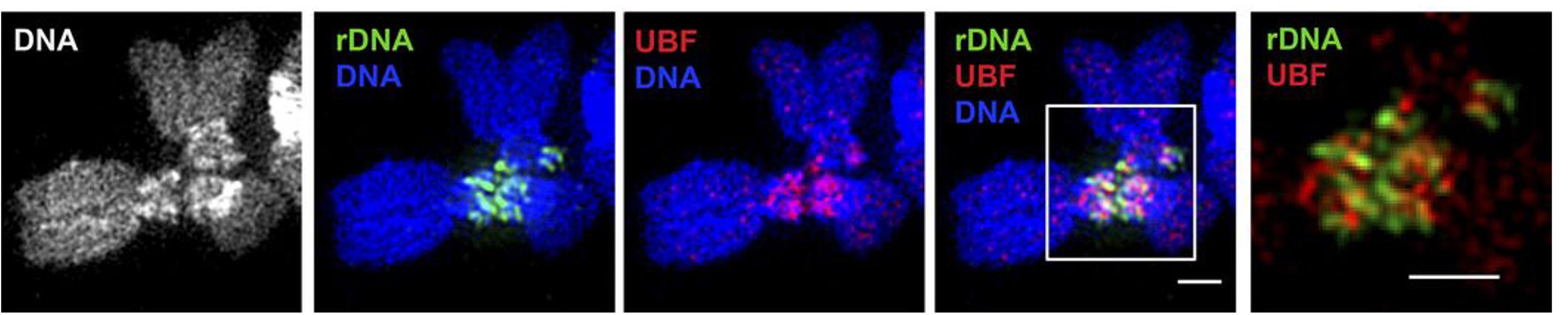
assembled between homologous sister pairs. SIM has also been used to
study a common core configuration of essential centromeric components in
human cells by Investigator Jennifer Gerton and her colleagues.