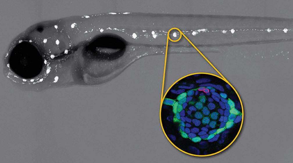
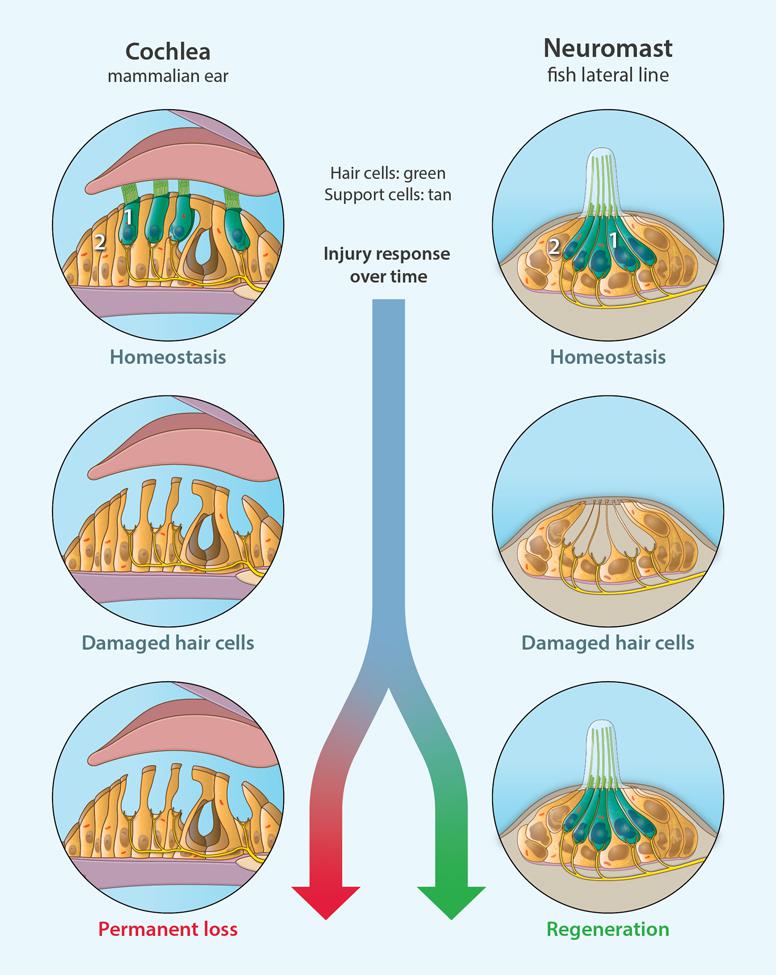
“The system is really ideal to study regeneration because sensory hair cells in the zebrafish lateral line regenerate extremely fast. This is one of the fastest regenerating cell types in a vertebrate described so far,” said Piotrowski.
Although an organ is composed of different cell types, the DNA in each of these cells is identical. What differentiates cell type, or cell gene expression, are the specific DNA sequences, or genes, that are transcribed into RNA messages. In this study, researchers use a technique called single cell RNA sequencing (scRNA-seq) to identify the activity of genes and cell types in HC regeneration at six, closely spaced time points.
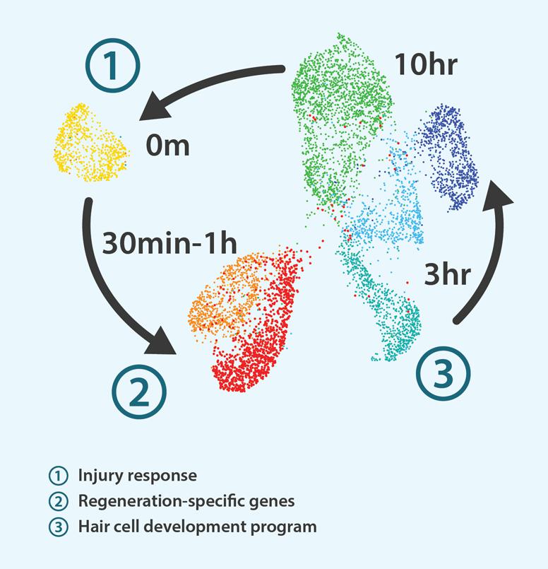
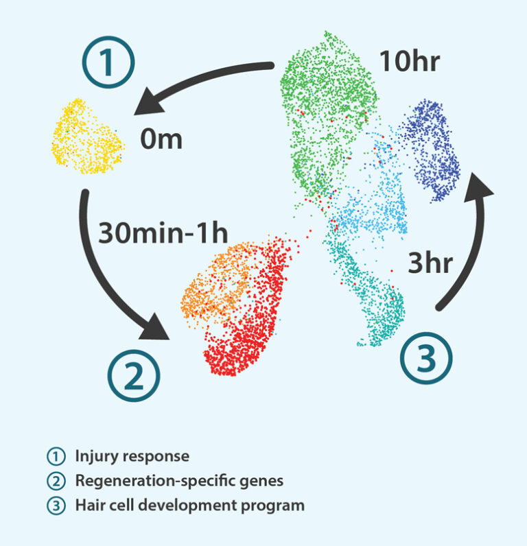
Analyzing gene expression just minutes after inducing HC death and at five additional time points up to 10 hours reveals an exciting discovery; gene activity in SCs and in HC progenitors—a type of cell similar to a stem cell that maintains developmental gene programs—falls neatly into three unique time periods or modules where the preceding module activates the next one.
In the first module (0-30 minutes), immediately following HC death, many homeostatic genes are turned off while “injury response” genes are briefly activated. This time frame allows SCs to essentially “reprogram” their function and fate, and, would have gone unnoticed had researchers not collected data right away.
Between 30 minutes and one hour, or in module 2, while many gene activities are still inhibited, some genes referred to as “regeneration-specific” genes are temporarily activated. “What is interesting about these regeneration specific genes is that several of them are described in fin, retina and heart regeneration,” said Piotrowski. “It could well be that some of these genes or pathways are conserved between different organs.”
In the third module (3-10 hours) starting approximately three hours post HC death, researchers find a sudden upswing and reactivation of the programs that allow cells to regenerate. “We see a lot of genes suddenly turn on that are involved in transcription and protein translation that you would need to make hair cells during development,” said Piotrowski.
Another interesting discovery is that only a small subset of SCs actually become HCs with the rest returning to their original state, a decision occurring between three and five hours. “Somehow the cell senses that it has enough hair cells and suppresses further hair cell fate.”
“The big question now is can we also find these three modules when we look at other regenerating organs,” said Piotrowski.
The researchers believe the initial inflammatory response activates regeneration but then needs to be suppressed for regeneration to proceed. The inhibition and subsequent enhancement of specific homeostatic genes is equally important; “downregulated” genes potentially could be temporarily turned off in mammals to aid in HC regeneration.
“The timing aspect of regeneration has not been described well so far,” said Piotrowski. “Knowing when to turn genes on and off and in which order can improve our understanding.”
The Hearing Health Foundation, of which Piotrowski is a member, aims to enhance our understanding of hearing loss via collaborative research to discover a cure. All data from the study is publicly available with the hope that scientists studying hearing loss and regeneration can use this to perform comparative studies across different organs and species including mammals.
“Our work provides a preliminary atlas of cell type and gene expression profiles. We hope to identify which genes are required to activate each module and to set a foundation for future studies focused on regeneration,” said Piotrowski.
Authors include Sungmin Baek, PhD, Nhung T. T. Tran, PhD, Daniel Diaz, Ya-Yin Tsai, Joaquín Navajas Acedo, PhD, Mark E. Lush, PhD, and Tatjana Piotrowski, PhD. This work was funded by the National Institute on Deafness and Other Communication Disorders of the National Institutes for Health (award 1R01DC015488-01A1) and by institutional support from the Stowers Institute for Medical Research. Data visualization and dissemination via gEAR was supported by grant R01DC019370.