News


22 July 2026
Coral Keepers
Inside our coral spawning success. In the heart of the midwest.
Read Article
News
“I was always so fascinated by those images,” says Peloggia de Castro. “I looked at them and thought, one day I want to do that."

By Marla Broadfoot
When Julia Peloggia de Castro was working in a biochemistry lab as an undergrad at the University of São Paulo, her boss used to tape pages from Nikon’s Small World calendar over every bench. One image featured a close-up of a bee eye dotted with dandelion pollen grains. Another highlighted individual nerve fibers in an array of pastel colors. Yet another zeroed in on a snowball of mold accumulating on an overripe tomato.
“I was always so fascinated by those images,” says Peloggia de Castro. “I looked at them and thought, one day I want to do that. I want to be the person who takes an image that will inspire someone else.”
Then in 2017, while interviewing for the PhD program of the Graduate School of the Stowers Institute, Peloggia de Castro received a colorful calendar showcasing winners of an Institute-wide scientific image competition. Her reaction? “Oh yeah.” She promised herself that she would participate in the image competition every year, no matter what.
At Stowers, incorporating scientific images into an annual calendar is just one way to explore the intersection of science and art. By commissioning illustrations and artwork, supporting artists-in-residence, and creating library and art museum exhibits, the Institute is helping people see science through a different lens, celebrate its accomplishments, question its assumptions, and understand its impact.
These efforts ultimately demonstrate that scientists and artists are not all that different. Both seek to create something new, to better the understanding of life, and to depict some version of reality. Their work—regardless of whether it comes from a laboratory or a studio—shows that truth and beauty can coexist.
An homage to science
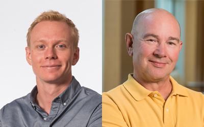
“Some people tend to picture scientists as white males in lab coats with pencils sticking out of their pockets who think about science all the time,” says Robb Krumlauf, PhD, an investigator and scientific director emeritus at Stowers. “In fact, many scientists come from diverse backgrounds, and many scientists have diverse interests.”
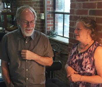

Robb Krumlauf, PhD and Kathy Barnard
Krumlauf says that though he possesses no artistic skills himself, he has always appreciated art and has been friends with artists throughout his life. He loves to visit local galleries when he travels around the world to scientific conferences to present his groundbreaking work on developmental biology. In 2016, Krumlauf was elected to the National Academy of Sciences, considered to be one of the highest honors given to a scientist in the United States. To celebrate the occasion, his wife, Leanne Wiedemann, commissioned their friend and Kansas City artist Kathy Barnard to create a piece of stained-glass art.
For inspiration, Barnard visited Stowers, where she learned that Krumlauf’s work is as philosophical as it is scientific. His research revolves around one central question—how can the creatures roaming this earth come in such a wide variety of sizes, shapes, and forms when their underlying genetic makeup is often so similar? Outwardly, fruit flies and humans could not seem more different. Yet they share a whopping 60% of their genomes. A mere 2.5% of DNA separates mice and men.
Krumlauf discovered that a set of genes called homeobox genes appear again and again in evolution, determining how the patterns of various tissues and structures unfold to give rise to each unique living and breathing organism. In fruit flies, mutations in these genes can create an extra pair of wings or prompt a leg to grow in place of an antenna. In mice, alterations in these genes can generate new pairs of ribs.
During Barnard’s visit, she noticed a delicate little pen-and-ink drawing of a mouse with wings hanging on the wall of Krumlauf’s office. The picture was a gift from Rosa Beddington, a friend and fellow scientist, who was inspired by Krumlauf’s work to draw the fanciful creature.
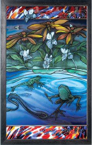
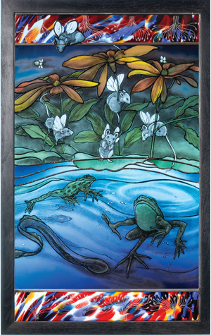
“I knew I had to start with that mouse with wings,” says Barnard, “but what else could I put in it? What kind of story could I tell in this piece of glass?”
Then Krumlauf explained to her that even though he began his research in mice, over the last several decades he had expanded his studies to include other experimental models like zebrafish, African frogs, and lamprey. In doing so, his studies have uncovered how the mechanisms controlling brain development differ between species, and how they can be disrupted in human diseases.
“I decided to incorporate all of these animals into the piece and have them interact to make the piece come alive,” says Barnard. The resulting piece features some playful mice with wings, frogs, fish and a lamprey swimming in the pond, along with a few insects buzzing around the flowers of an echinacea plant. The borders are etched with patterns reminiscent of the nerves branching out of the brain region Krumlauf studies. At the top of the piece, a curious mouse peers into the scene below. “I included that element to bring you in,” says Barnard. “You are that mouse, observing it all.”
Early in the process, Krumlauf visited Barnard in her studio to observe the artist at work. He watched as Barnard employed chemistry, heat, and color to immortalize his model organisms in glass, using a technique she had honed over the years. “I loved seeing how she developed things, how it all came together,” says Krumlauf. “It does remind me of how we as scientists make discoveries. It involves a lot of practice, and trial and error. Sometimes accidents are made. Sometimes it turns out beautifully.”
Today, the stained-glass piece—which turned out beautifully—hangs in Krumlauf’s office at Stowers. Sometimes, like when it catches the midafternoon light, Krumlauf says he finds it hard not to daydream. “To me it is a celebration of creativity and discovery for both art and science.”
Science simplified
While science often inspires art, as it did when Barnard etched a menagerie of model organisms into stained glass, art can also inform science. Such is the case with Darrick Hansen, a predoctoral researcher working in Linheng Li’s lab at Stowers. Hansen has been studying adult stem cells that reside deep inside the gut, in millions of small pits or “crypts.” These amazing regenerative cells respond to a multitude of signals from the environment and other cells in the crypt as they rebuild the entire lining of the gut, a feat they perform every few days.
“There ends up being so many players that using words alone to describe them starts to feel impossible,” says Hansen. Li suggested that Hansen reach out to Mark Miller, a science and medical illustrator at Stowers, to create a visual depicting the “wild, wild West” of the intestinal stem cell niche. “Though there are lots of images out there already, they aren’t very aesthetic,” says Hansen. “And they don’t give a good sense of what is going on in this complex structure.”

Darrick Hansen and Mark Miller
The duo began meeting in 2017 to develop the illustration, which they wanted to serve as a living resource that could be built upon as knowledge grew. Miller started out with what was known about the structure historically, drawing upon histology and anatomical studies of the architecture of the niche. Then he brought in the new data Hansen had gathered about the types of cells that had been discovered lurking in the specialized space.
As Miller reconstructed the scene, it became clear where gaps in his own knowledge—and sometimes that of the field—remained. He began peppering Hansen with questions. How big is this cell type? How far away is it from that structure? Where are the blood vessels? “Some of these questions might be considered ignorant, but I needed to answer them to create the illustration, as it’s really difficult to draw what you don’t understand.”
Hansen says those questions forced him to look at the data in greater detail, and to question some of the assumptions in the field. “When you are always talking to the same people, you assume certain things,” he says. “But then when someone from the outside asks a question, you say, wait a second, is there data to support that? Or is that just something that we pass down anecdotally or a term that we’ve used incorrectly all this time?”
After nearly two and a half years of back-and-forth discussions, the team developed an illustration that is complex, brightly colored, and, not surprisingly, difficult to put into words. At first glance, the looping, invaginated forms of the crypts might resemble a roller coaster running on a network of circuits embedded underneath. But with closer inspection, the interactions between 15 different types of cell types begin to emerge.
Hansen says his conversations with Miller have helped him grow as a scientist. He says he is no longer afraid to ask questions or to risk appearing stupid in his quest for the truth. “I think presumed knowledge causes far more problems in science today than ignorance does,” he says.
Though the illustration has not been published yet, Hansen has shared it with other members of the NIH-funded Intestinal Stem Cell Consortium, of which the Li Lab is a member. He says, while several researchers have commented about learning something new, a couple have pointed out something they thought was wrong.
“I said no, that’s right,” says Hansen. “Look at the data.”
Worth a thousand words
While scientific images convey valuable data to researchers, their simple beauty can sometimes transcend the information they contain and transform them into objects of art. Once a year, Crossroads, an organization of postdocs, students, and staff scientists, asks researchers to enter their best photographs, data visualizations, and videos into a scientific image competition held as part of the annual Young Investigator Science Retreat (YISR).
“We set up Crossroads to help advance the careers of young scientists, to help them develop the soft skills they need to succeed,” says Krumlauf, who oversaw the program when he was Institute director. “But one of the things we discovered when talking with trainees was how proud they were of the data coming off their microscopes or the drawings they created on their computers. You would see their work framed in their offices or posted in the hallways.”

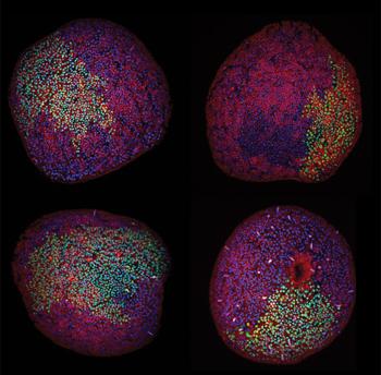
Krumlauf decided to compile Stowers researchers’ most visually appealing work in one place, by showcasing the YISR images in its “Images of Discovery” calendar. The calendar features not only photographs but also intricate illustrations and data visualizations created by trainees to communicate their science. “It’s the whole idea of a picture being worth a thousand words,” he says.
One image, reminiscent of a busy map of airport hubs, actually depicts a constellation network of gene expression in Mexican cavefish and surface fish. Another that looks like orange and yellow pinwheels is in fact a series of rose plots representing the number of Stowers-affiliated publications per month over the course of four years.
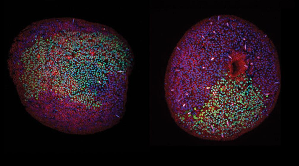
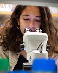
Stowers Predoc Peloggia de Castro made it into last year’s calendar with an image that features four bright red and blue balls of cells covered in smatterings of green, showing the starlet sea anemone at early stages of development. When she traveled home to São Paulo, she took a handful of copies of the calendar with her to share with her grandmas. The calendar not only gave them bragging rights but also served as a conversation piece for her to talk to people in her community about science.
“My family loves it,” she says. “They don’t understand anything that is written there, but they always leave that month up all year long. When anyone came to visit the house, they would see the picture and I would explain to them what I do. It makes me really happy because it helps me connect with people who are not scientists.”
In the eye of the beholder
Microscopists Jay Unruh, PhD, and Brian Slaughter, PhD, both grew up on farms in the heartland, where the houses were far apart and there were no scientists as far as the eye could see. Yet they both loved science, and visited their neighborhood libraries to pick up fun facts about biology and physics or pore over biographies of famous inventors. Today, as co-directors of the Stowers Microscopy Center, they help organize events at local Kansas City libraries to expose the public to science and the scientists that conduct it.
“We were raised in communities that didn’t necessarily know or trust scientists,” says Slaughter. “We think it is important for the public and scientists to interact as much as possible, and we use art as a way to do that.”
For the “Beauty of Biology” exhibit, the pair curated some of the Institute’s most eye-catching images—many winners of the image competition—and printed them onto large sheets of high-gloss metal. They then arranged the images into a series, first depicting whole organisms, then parts of organisms, and finally cells and structures that are invisible to the naked eye. Unruh said the exhibit was designed to draw people in, to wonder and to ask questions about the meaning underlying the beauty.
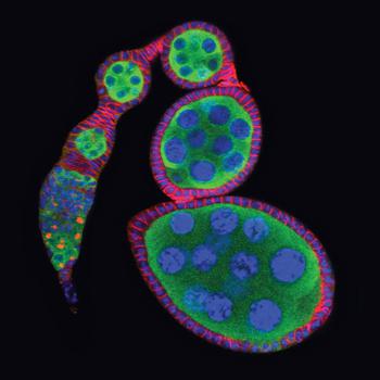
Dani Wellemeyer, head of outreach and community engagement at the Miller Nichols Library at the University of Missouri-Kansas City (UMKC), recalls being captivated by the larger-than-life views of biology afforded by the traveling exhibit. Her favorite image depicted the ovaries of a fruit fly as a cobblestone path of green blobs, each outlined with red and blue hatch marks and filled with bubbles of blue material. Wellemeyer was not the only one picking favorites; people often asked her if they could buy copies of the images to take home with them.
In addition to the Miller Nichols Library, the exhibit has made the rounds to four community libraries, each for several weeks to a few months at a time. At most of the libraries, “show-and-tell” receptions were held, featuring Stowers research trainees to work microscopes they brought for the occasion or to answer questions from the attendees.
“The big questions that we always try to prepare our students for are ‘Why do you care about what happens in a flatworm or a sea anemone or a mouse? How does this relate to human health?,’” says Unruh. “I think those are hard questions for students to answer, but they are important.”
Through the process, the organizers found that they often disagreed with members of the public about what was beautiful about the works on display. One time, they heard some people commenting on the juxtaposition of vibrant reds, yellows, and greens in a particular image, and all Unruh and Slaughter could see were the underlying scientific ideas that it portrayed.
“We’re thinking about the cool processes of science that are represented here, that we don’t completely understand,” says Slaughter. “And we can think of all the fun experiments we’d like to try to understand them. We think it is beautiful, but from a different perspective.”
Bridging disciplines
Science prizes objectivity, while art works to the opposite effect. Still, both can tickle the intellect and stir the emotions. Labeling science and art as separate entities belies the fact that these two drivers of human innovation have been intertwined for centuries, in the mind’s eye as well as on the world stage. The epitome of this interplay is the Renaissance artist and scientist Leonardo da Vinci, whose studies of anatomy and physiology brought the Mona Lisa to life.
Steph Nowotarski, PhD, a postdoc in Alejandro Sánchez Alvarado’s lab at Stowers, considers herself to be both an artist and a scientist. She went to college planning to double major in her two loves but quickly found there were not enough hours in the day for her to spend in the lab training to be a scientist and to spend in the studio training to be an artist. “As society accumulates knowledge at an exponential rate, we are asking people to learn things very quickly, and so you have to stay in your lane to keep up,” she says.

Mol Mir
Though Nowotarski made what she considered the pragmatic choice to stay in the science lane, she kept looking for opportunities to veer into art. One day she met Mol Mir, a student at the time at the Kansas City Art Institute (KCAI) who was touring Stowers with classmates. Mir asked an insightful question about how the detector worked on the high-powered microscope Nowotarski was using. That question led to a conversation about how Mir’s art focused on generating 3D renderings of cells, a technique Nowotarski had just begun to explore for her own work on the planarian flatworm. That summer, Mir joined the Sanchez lab as Stowers’ first art intern, later staying on as a lab assistant.
“The science that I do mostly involves looking at electron micrographs of planaria and building models off of them,” says Mir. “So my art is constantly inspired by what I am seeing every day.”
Mir and Nowotarski teamed up with retired KCAI fiber chair Jason Pollen and recent KCAI graduate William Plummer to launch an immersive art exhibit inspired by planaria, a model organism made famous by its regenerative properties. The team met once a week to brainstorm ideas and play around with different mediums and experiences, all exploring what the minuscule aquatic creatures could teach us about being human.
The exhibit, which was displayed at the UMKC Gallery of Art from January through early March 2020, took viewers through two separate rooms—one dark, one light. The dark room explored the concept of observation, the first step in any scientific or artistic process. Viewers strolled past giant projections of planaria, blown up hundreds of times their actual size. They saw photos of the arrow-shaped flatworm cut into pieces, alongside photos after these pieces morphed into full-sized clones. They were captivated by the 16-minute video A Drawn Line, in which the animals undulated and moved with perfect symmetry, as if trapped in a kaleidoscope.
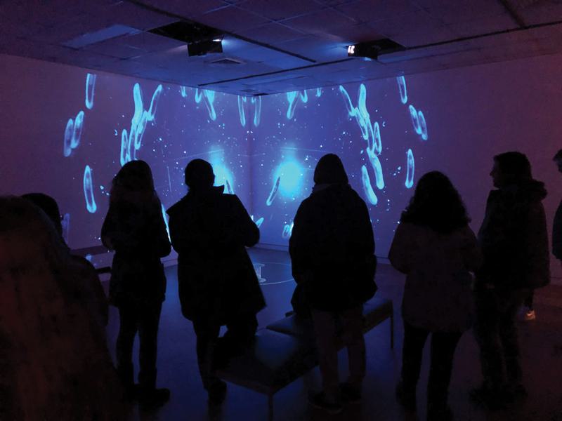

In the second room, people were treated to wall-to-wall canvases featuring interpretive works of art by Mir, Nowotarski, Pollen, and Plummer. Each of these static paintings evoked a sense of movement, either by a maze, a mass of overlapping circles, or an elaborate mandala. In the center of the room, a single pendant light hung over three cylindrical tanks displaying the planaria. The tanks were perched on a table crafted in the shape of a planaria cut into three pieces, left over from Mir’s thesis project. The room had plenty of space for people to interact and interpret the artwork, and peer at the animals with magnifying glasses, marveling at how small they were in real life.
“Science and art can both have a lot of jargon. I was excited to see scientists asking questions about art, and artists asking about science,” says Nowotarski. She said the experience taught her there are many ways to communicate one’s place in the world. And to remember to tap the same creativity she channels through her art to fuel her pursuit of science.
Truth and beauty
Science and art both take creativity, curiosity, and persistence. These seemingly disparate disciplines emphasize the importance of observation and interpretation, often leading to big ideas that force us to shift our worldview. It is hard to imagine where we would be today without the knowledge gained by science or the inspiration gained by the arts.
Peloggia de Castro, who has spent two years working as a predoctoral researcher in Tatjana Piotrowski’s lab, hopes to have a lab of her own one day. But she worries if that will be possible, given the ever-shrinking number of tenure-track academic jobs available. On bad days, when the failures that are a regular part of the scientific process get her down, she waits until everyone has left before firing up the microscope to take beautiful pictures.
There, she takes close-ups of phenomena most of us will never see, much less appreciate. The tiny heart of a growing zebrafish, whose body is almost entirely transparent. The migration of a hundred specialized cells from the head of that remarkable organism all the way to the tip of its tail. The “zebrabow,” whose cells light up with the colors of the rainbow.
After a couple of hours lost in that hidden world, Peloggia de Castro remembers why she went into science. “Life is beautiful,” she says. “This is beautiful. This is what I fell in love with.”
News
22 July 2026
Inside our coral spawning success. In the heart of the midwest.
Read Article
News
22 July 2026
Dive into the biological basics of symbiosis, the close partnerships that allow different organisms to thrive together, and take a closer look at how a Stowers scientist explores the molecular conversations that make these relationships possible.
Read Article
News


17 July 2026
Evan Morrison, Ph.D., a Stowers Institute postdoctoral researcher and a 2021 recipient of the Howard Hughes Medical Institute Gilliam Fellowship, followed an unconventional path into biological research, and continues his HHMI fellowship at the Stowers Institute researching how cells identify defective ribosomes.
Read Article


