Understanding an organism’s “cellular catalog”—the classification of the various types and numbers of cells it possesses—reveals much about how it thrives, dies, or otherwise responds to changes in its environment. For instance, identifying the types and amounts of immune cells in diverse animals can help researchers understand their particular immune system responses as well as immune system adaptation to different environments.
To determine the cellular catalog of an organism or a tissue, researchers often use cell imaging techniques to identify observable changes in cell populations. But these tools historically are useful only for organisms that have a long history of scientific literature on well-recognized cellular characteristics, such as mice, fruit flies, zebrafish, or even humans.
What if the goal is to study the composition of cells in organisms that have yet to be fully characterized? In a recently published paper, a collaborative team of researchers from the laboratories of two principal investigators and two technology centers at the Stowers Institute for Medical Research describe a new image-based cell classification tool.
Known as Image-Cytometry Cell Classification, or Image3C, the tool allows researchers to characterize the cell make-up of tissues at single-cell resolution in research organisms when pre-existing knowledge about their cell types is not available. Image3C uses intrinsic cellular features and broadly-acting dyes to perform this cell composition analysis.
The tool clusters cells based on their appearance to reveal groups that represent different parts of tissues and organs, providing previously inaccessible clues as to how tissues and organs develop and function. Image3C also allows detection of changes in cells subjected to different conditions to allow researchers to investigate the impact of environmental factors on cells and tissues.
Classifying cells in less-studied research organisms is now easier, thanks to a new tool developed at the Stowers Institute.
Robert Peuß, PhD
Intramural team
Origins of the project began in 2016 when Robert Peuß, PhD, then a postdoctoral research associate in the Stowers Institute’s laboratory of Nicolas Rohner, PhD, was studying the immune system of cavefish—descendants of river fish swept into dark underground caves by floods thousands of years ago.
“I found we were not able to use conventional methods to understand the immune cells of these cavefish,” said Peuß. “There just was not a good way to enumerate cells from different tissues in emerging research species like the cavefish because they don’t have a large molecular toolkit yet.”
When he first joined the Rohner Lab, Peuß’s mentor encouraged him to use the technology centers at the Institute and the expertise of their teams as much as possible because they could help establish methods for studying cavefish that could be used for the project at hand as well as for the entire Institute.
Andrew Box
Following this guidance, Peuß reached out to Andrew Box of the Cytometry Facility at the Institute who had the idea of using a new flow cytometry instrument called the ImageStream to take many images of single cells within the cavefish. Standard flow cytometry measures physical and molecular characteristics of cells suspended in a liquid that flow in single file by lasers and emit signals captured by detectors. The ImageStream adds high-quality imaging to flow cytometry, allowing the capture of many detailed images per sample to reveal additional features of the cells.
“We ran the cavefish samples on this imaging flow cytometer and were really impressed with the images that we found,” said Box. The team quickly realized this approach likely had broader applications with other types of automated or high-throughput analysis approaches.
Given the collaborative nature of research at the Institute, the duo sought the expertise of others who might also be interested in this new kind of single-cell imaging capability.
“Many at the Stowers Institute work with research organisms that have not been used extensively in the field and therefore can’t be studied with conventional methods,” said Peuß. “We wanted to work together with as many labs as we could to make a tool that is useful for the entire community of people who work on non-classical research organisms at the Institute.”
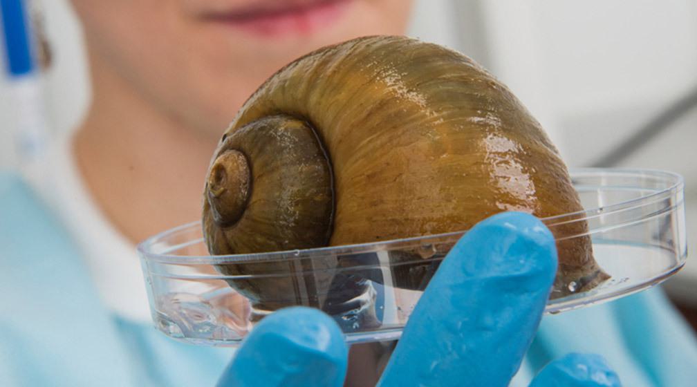
“I also needed tools to answer biological questions and I had talked with individuals in the technology centers for suggestions and techniques to help me figure them out,” Accorsi recalled. Box immediately thought of his project with Peuß and suggested they use the newly developed Image3C on her apple snails.
“We wanted to know more about the cellular composition of these organisms, so our labs joined forces,” Accorsi said. “It was a great opportunity to combine our expertise and draw on the technology centers at the Institute to make a global tool for all kinds of different organisms and tissues.”
Neural network trains the tool
Building out the rest of the Image3C team was Chris Wood, PhD, of the Institute’s Microscopy Center who introduced the idea of using a kind of artificial intelligence called deep learning in the analysis pipeline. Specifically, Wood worked on a convolutional neural network approach which uses layers of mathematical operations and algorithms to detect visual aspects in the images that may be undetectable by human observation. Notably, the convolutional neural network carries out its detection analysis in an unbiased way.
Chris Wood, PhD
“When Chris joined the project, he brought it to the next level beyond clustering cells by size and groups as we did at first,” Accorsi said. Wood’s expertise helped overcome a major problem of the first iteration of the tool: it required a lot of time-consuming re-computing. Every new set of samples needed to run through the workflow.
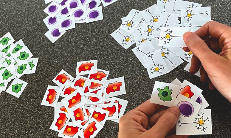
“That’s where the neural network became really important because once we ran through a representative data set, we could train it to understand different cells within the organism,” Wood explained. The neural network can learn certain cell image aspects and then classify them, a reason why the team refers to Image3C as a “cell classifier machine.” Think of it like a person viewing photos of several dogs and classifying them into breeds based on visual characteristics, but with greater accuracy and less bias.
By training the machine with several samples, the tool became an even faster high-throughput device, speeding up the cell grouping process with less re-analysis and hands-on time. It also made analysis of follow-up experiments much easier by lowering the risk of bias compared to manual cell counting.
Biology validated the computation
To check the validity of the new tool, the team established a quality control effort with known samples to be sure the output matched expected results.
Nicolas Rohner, PhD
“It was a really nice back-and-forth between Andrew and Chris on the technology side, and Rob and I on the biology side,” Accorsi recalled. As Box and Wood were writing the script, running the samples on the tool, and sending the results to the labs, Peuß and Accorsi reviewed the data from a biological perspective using both a well-studied organism (zebrafish) and a less-studied one (apple snail). “It was an excellent integration of tool development and double-checking the output to see if it was making sense biologically.”
Scientific freedom
Peuß and Accorsi credited the scientific freedom the Institute provides for making the project happen. They mentioned that both the Rohner and Sánchez Alvarado laboratories gave them as postdocs scientific latitude and resources to pursue compelling new research.
“There is a lot of support to be brave about trying new things and exploring new concepts,” said Peuß.
Sánchez Alvarado agreed. “At the Stowers Institute, we expect our postdoctoral scientists to be both independent thinkers and mentors. When Robert and Alice discussed their idea with us, we did not hesitate to support their effort. Often, it is by following the trail of curiosity that scientists acquire new and transformative insights.”
Alejandro Sánchez Alvardo, PhD
The Stowers Institute research environment encourages its teams to take risks, explore new approaches, and take the time needed without knowing exactly whether a project will work or not.
“There are many opportunities and situations that bring people from the research labs and the technology centers together,” said Accorsi. “It is so appreciated to have these opportunities to start conversations that often lead to common ground where we can work together.”
For the Image3C team, knocking on the doors of colleagues not only helped solve their own research questions of the day but strengthened capabilities for broader use at the Institute and elsewhere.
“Existing tools were difficult to use for less-studied research organisms,” said Peuß, who is now at the University of Münster, Germany. “Our tool is really filling a gap at the Institute and indeed in the larger research community of scientists who are working with uncommon organisms.”