News

22 June 2022
The switch-like nature of the immune system
Immune cells respond to danger in an “all-or-none” fashion via protein aggregation
Read Article
Press Release
Stowers scientists deduce the initiating structure of the amyloid implicated in Huntington’s and present a potential therapeutic treatment approach.
Randal Halfmann, Ph.D., discusses new research revealing the start of Huntington's disease
KANSAS CITY, MO—June 13, 2023—Devastating neurodegenerative diseases like Huntington’s, Alzheimer’s, and Parkinson’s are all associated with protein deposits in the brain, known as amyloid. Despite extensive research investment into the cause and toxicity of amyloids, deciphering the first step in formation along with effective therapies has remained elusive.
For the first time, scientists at the Stowers Institute for Medical Research have uncovered the structure of the first step in amyloid formation, called the nucleus, for Huntington’s disease. The study published in eLife on June 13, 2023, from the lab of Associate Investigator Randal Halfmann, Ph.D., proposes a new, radical method for treating not only Huntington’s but potentially dozens of other amyloid-associated diseases—preventing the initial, rate-limiting step from occurring.
“This is the first time anyone has experimentally determined the structure of an amyloid nucleus even though most major neurodegenerative diseases are associated with amyloids,” said Halfmann. “One of the big mysteries of Huntington’s, Alzheimer’s, and ALS is why disease coincides with amyloid, yet the amyloids themselves are not the main culprits.”

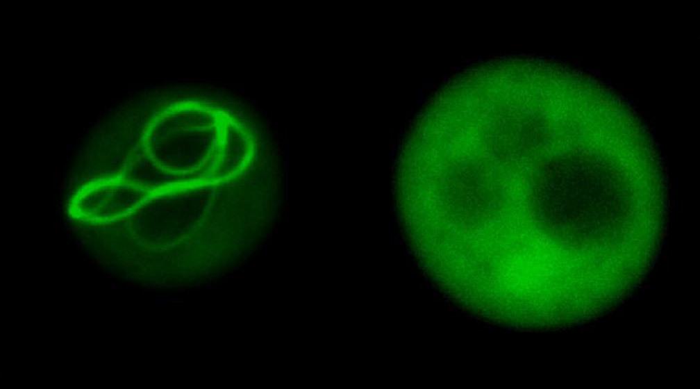
Fluorescence lifetime micrograph of a fluorescently tagged human protein inside yeast cells. Different colors indicate different states of protein aggregation.
Co-first authors Tej Kandola, Ph.D., and Shriram Venkatesan, Ph.D., uniquely identified the structure of the amyloid nucleus for huntingtin, the protein responsible for Huntington’s disease, discovering that the nucleus forms within a single protein molecule.
Proteins are the cell’s factory workers constructed from unique sequences of 20 amino acids, their building blocks. Some proteins have repeats of one of these amino acids—glutamine (abbreviated as Q). Huntington’s and eight other diseases, collectively called “PolyQ diseases,” occur when certain proteins have a repeat that is too long. Somehow, this causes the proteins to fold into a specific structure that starts a chain reaction that kills the cell.
“For three decades, we’ve known that Huntington’s and related fatal diseases occur when proteins contain more than around 36 Qs in a row, causing them to form chains of proteins in the brain, but we didn’t know why,” said Halfmann. “We’ve now figured out what the first link in the chain looks like, and, in doing so, have discovered a new way to stop it.”
“I am, frankly, astonished that such an intuitive physical model of nucleation emerged despite the intrinsic complexity of the cellular environment,” said Professor Jeremy Schmit, Ph.D., from Kansas State University. “I am truly excited by the intuition and the testable hypotheses that this work inspires.”
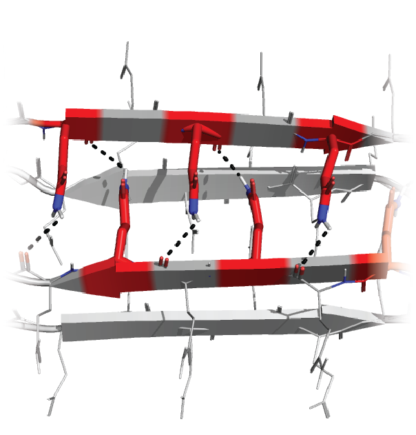
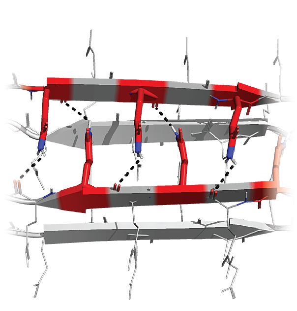
Graphical illustration of the polyQ nucleus. Glutamines (Q) interdigitate between two antiparallel two-stranded beta sheets.
A paradigm shift and potential therapeutic method
These new findings are potentially a paradigm shift for how we view amyloids. The results from this research suggest that it is the early committed steps of amyloid formation, right after the nucleus forms, that cause neuronal cell death.
Along with uncovering the key structure that begins polyQ amyloid formation, researchers found that it only formed in isolated molecules of the protein. Clumping the proteins together in cells stopped amyloids forming altogether. This is a novel therapeutic avenue the team plans to explore further in mice and brain organoids.
A new technique
A technique recently developed by the Halfmann Lab, Distributed Amphifluoric Förster Resonance Energy Transfer (DAmFRET), shows how a protein self-assembles in single cells. This method turned out to be crucial for observing the rate-limiting amyloid-forming nucleation event.
“A key innovation was to minimize the volume of the reaction to such an extent that we can witness its stochasticity, or randomness, and then we tweak the sequence to figure out what is governing that,” said Halfmann.
Designing and testing specific patterns of Qs enabled the team to deduce the minimum structure that could form amyloid—a bundle of four strands each with three Qs in specific locations. This tiny crystal inside a single molecule of the protein is the first step in a chain reaction that results in disease.
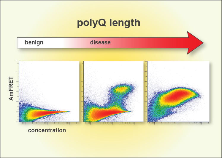

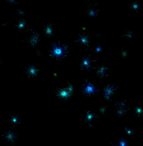
DAmFret plots illustrating the extent of glutamine (Q) self-aggregation as the number of sequential Qs increases (left: Qs less than 36; center: Qs around 50; right: Qs over 100).
“Prior work in test tubes supports a monomeric nucleus, but this model has been controversial,” said Halfmann. “We now have strong evidence that 36 Qs is the critical number for nucleation to happen in single protein molecules, and moreover, that this is how it happens inside living cells.”
In essence, this work provides a molecular model to investigate the structure of any amyloid nucleus. Additionally, the correlation between aging and amyloids suggests that this method may ultimately uncover molecular mechanisms that cause aging. The preemptive approach to eliminate or at the very least to delay nucleation provides hope for people with pathologic PolyQ proteins.
“The emerging paradigm is that everything follows from a single event, a spontaneous change in protein shape,” said Halfmann. “That event ignites the chain reaction for amyloids that kill cells and may provide critical insight into how amyloids cause disease.”
Additional authors include Jiahui Zhang, Ph.D., Brooklyn Lerbakken, Alex Von Schulze, Ph.D., Jillian F Blanck, Jianzheng Wu, Ph.D., Jay Unruh, Ph.D., Paula Berry, Jeffery Lange, Ph.D., Andrew Box, Malcolm Cook, and Celeste Sagui, Ph.D.
This work was funded by the National Institute of General Medical Sciences of the National Institutes of Health (NIH) (award: R01GM130927) and by institutional support from the Stowers Institute for Medical Research. The content is solely the responsibility of the authors and does not necessarily represent the official views of the NIH.
About the Stowers Institute for Medical Research
Founded in 1994 through the generosity of Jim Stowers, founder of American Century Investments, and his wife, Virginia, the Stowers Institute for Medical Research is a non-profit, biomedical research organization with a focus on foundational research. Its mission is to expand our understanding of the secrets of life and improve life’s quality through innovative approaches to the causes, treatment, and prevention of diseases.
The Institute consists of 20 independent research programs. Of the approximately 500 members, over 370 are scientific staff that include principal investigators, technology center directors, postdoctoral scientists, graduate students, and technical support staff. Learn more about the Institute at www.stowers.org and about its graduate program at www.stowers.org/gradschool.
Media Contact:
Joe Chiodo, Head of Media Relations
724.462.8529
press@stowers.org
News
22 June 2022
Immune cells respond to danger in an “all-or-none” fashion via protein aggregation
Read Article
In The News


03 January 2022
The Scientist
Read Article
Video
17 March 2023
As part of the #StowersImpact campaign, the Stowers Institute for Medical Research teamed up with the Alzheimer’s Association Heart of America and Greater Missouri Chapter to put a face to the disease that impacts so many people.
Read Article
Press Release
13 June 2023
How did ancient animals develop from egg to adult, and to what extent have the genetic mechanisms that guide embryonic development endured across millennia?
Read Article
