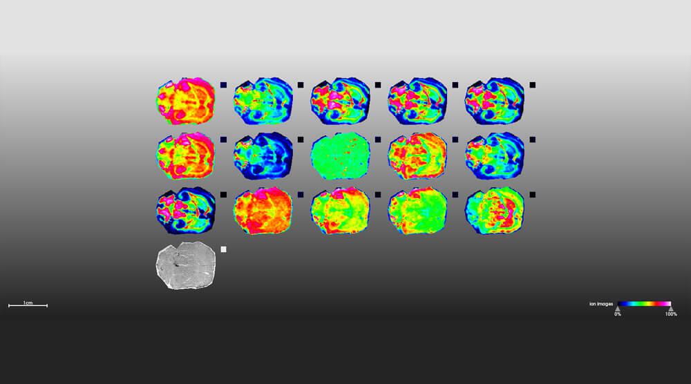
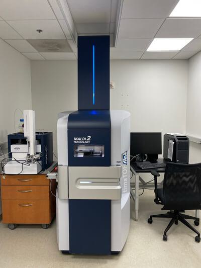
What does the timsTOF flex MALDI-2 do?
The timsTOF flex MALDI-2 can be used to identify a range of
biomolecules including lipids, metabolites, and proteins. In contrast to
our existing technology, the new instrument can analyze not only liquid
samples but also solid samples such as tissue sections. Consequently,
we can now map the distribution of these biomolecules across tissues at a
resolution of 50 micrometers, approximately the size of a grain of sand
or the width of a single strand of hair. This process is also referred
to as Imaging Mass Spectrometry.
What is the benefit of having this technology at Stowers?
The new technology will allow researchers to map the distribution of
large numbers of different biomolecules in tissues. For example, this
could allow identification of groups of biomolecules specific to
diseased tissue or subsets of biomolecules important for fundamental
biological processes such as regeneration.
What are you most looking forward to about having access to this technology?
As the name implies (“flex”), the new instrument is flexible. Using
ion mobility, this instrument is far more sensitive, allowing us to
characterize proteins from a single cell. Previously, protein analysis
by mass spectrometry relied on much larger samples prepared from
thousands of cells. This will open new avenues for research and will be
exciting to observe differences in the proteome from one cell to
another.
Can you describe a few collaborations that your team has utilized this technology in?
We are increasingly collaborating with the other Technology Centers
here at the Stowers Institute. For example, we coordinate with the
Histology team to prepare tissue sections on slides prior to MALDI
imaging. Single cell proteomics analyses involve collaboration with
Cytometry for cell sorting, and Automation for the robotics technology
needed for small-scale sample preparation.