#TechTalk: Q&A with Kevin Ferro, Scientist in Cytometry
Kevin Ferro is a scientist in the Stowers Institute’s Cytometry Technology Center.
30 January 2023
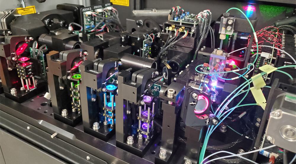
Interior of the ImageStream Mark II. Image courtesy of Kevin Ferro.
What exactly is cytometry?
Flow cytometry refers to the measurement of the molecular makeup of cells suspended in fluid. The single cell-containing fluid is passed through a set of lasers that, in combination with fluorescent probes specific to proteins or nucleic acids inside or on the surface of the cell, allow us to analyze and characterize large quantities of cells in real time based on their fluorescent fingerprint. This also enables us to isolate these cells for further experimentation or cultivation.
What do you think people would find most interesting about your job and the technology you use?
Many of the technologies we use to understand fundamental principles of biology require either extensive processing of samples, potentially altering the nature of cells and tissues studied, or are optimized to yield information about specific aspects, for example, the absence or presence of certain genes. Flow cytometry allows us to study life at the cellular level using live samples while measuring various characteristics at the same time, such as which proteins are expressed by the cell, its metabolic state as well as more general information about its morphology. This makes flow cytometry a key technology in the life and clinical sciences.
What does the ImageStream do?
The ImageStream Mark II is an image cytometer, a unique type of flow cytometer that combines the fidelity of fluorescence microscopes with the high-throughput capabilities of flow cytometers to allow detailed fluorescent images of large quantities of cells in suspension.
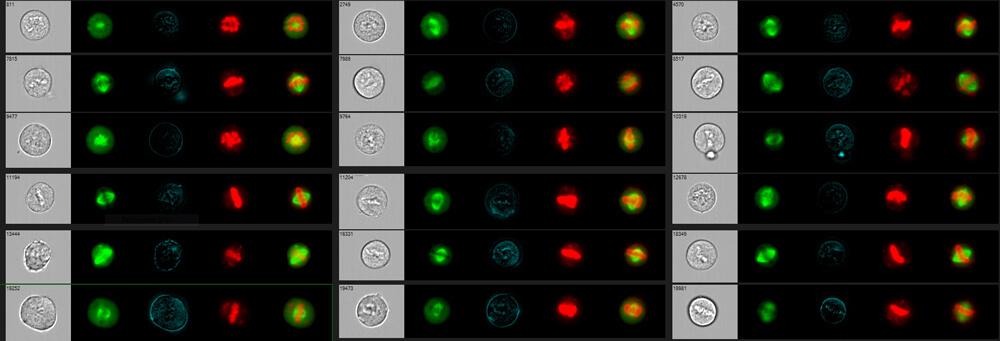
Human k562 in Metaphase expressing EGFP-beta-tubulin (green) and stained with a nuclear (red) and cell membrane (blue) dye. Image courtesy of Jillian Blanck and Bret Redwine.
What is the benefit of having this technology at Stowers?
At Stowers we often work with research organisms that are
understudied and therefore lack the tools established for more commonly
used systems such as mice or human cell lines. The ImageStream captures
large numbers of images of cells without the need for fluorescent probes
in order to study those cells using machine and deep learning
algorithms. This workflow facilitates the study of uncommon research
organisms and empowers our scientists to work with a more representative
array of species to answer fundamental questions about immunity,
genetics, development, and regeneration.
What are some of the most exciting discoveries made so far with this technology? Both at Stowers and elsewhere.
We recently published a machine and deep learning-based analysis method called Image3C
that featured novel insights into the immune systems of cavefish and
apple snails, two of the species studied at the Institute to better
understand metabolic dysregulation and soft tissue regeneration,
respectively.
Other areas of research that the ImageStream has had a large impact
on are small-particle biology as well as the study of rare circulating
leukemia cells, aging, and immune cell interactions. The most
important impact of image cytometry to me personally has been the push
to develop cell sorters powered by this technology. Various inroads to a
mature instrument have been made by research labs and companies alike
for several years now and access to image cytometry cell sorting would
revolutionize the way we study both well-studied and understudied
organisms here at Stowers and around the world.
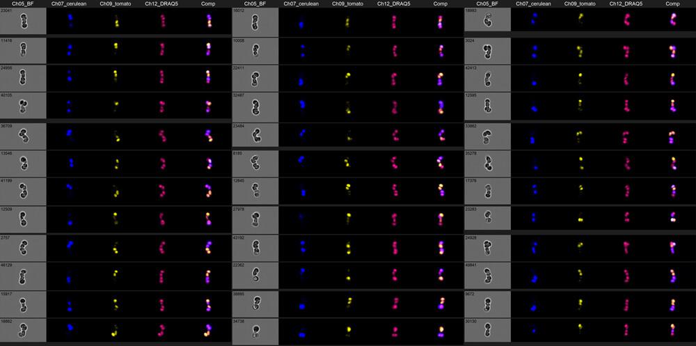
Fission yeast asci stained with nuclear dye (red) and expressing either the fluorescent proteins mCerulean (blue) or tdTomato (yellow-orange) in the spores contained in the asci. Image courtesy of Jillian Blanck.