For most animals, during development and in the maintenance of adult tissues, cells receive and respond to different sets of instructions that tell them where and what they should be. A given signal or cue initiates a series of steps that form a path from reception to response, known as a signaling pathway. Many of these pathways, when disrupted, can lead to diseases including many cancers.
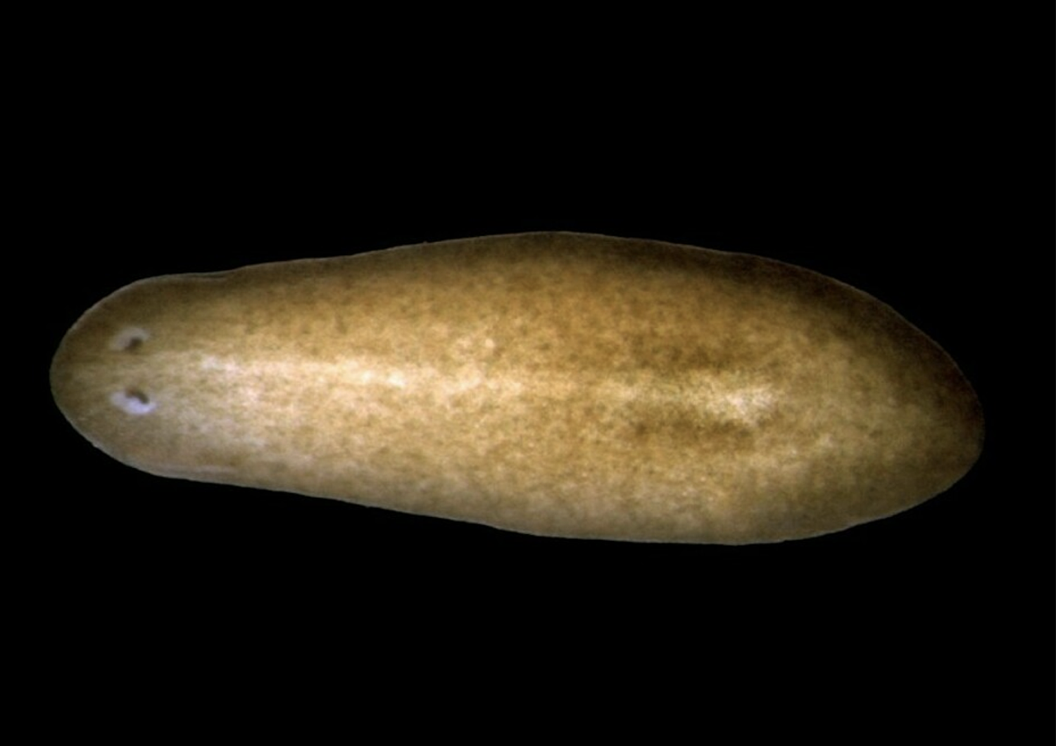
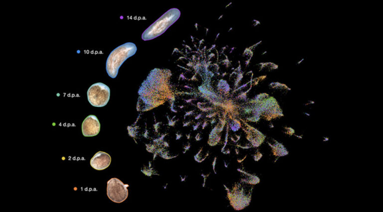
“Planarians can regenerate lost tissues and organs to form a fully functional animal in a span of two weeks,” said Viraj Doddihal, Ph.D., lead author of the study and a postgraduate researcher in the Sánchez Alvarado Lab. “These organisms have figured out a way to read information from the existing body while they rebuild tissues, similar to finding a location using GPS.”
“Our work describes a network of genes that coordinates the growth of new tissues so that the shape and size of the regenerated tissues and organs are proportional to the size of the animal,” said Sánchez Alvarado. “When missing a head, for instance, the animal employs this network to regrow a head of the right size in the right place.”
The study published in Proceedings of the National Academy of Sciences on May, 7, 2024, identified a protein, specifically an enzyme called pak1, that guides two different signaling pathways to coordinate proper patterning of two major body axes. In addition, the team found that pak1 interacts with components of another signaling pathway called Hippo. Thus, pak1 and the Hippo pathway form a genetic network that serves to direct body shape via axis patterning, enabling the pathways governing these two axes to communicate: one axis from head-to-tail and the other from center-to-edge. While in many animals the Hippo pathway is known to control organ size, in planarian flatworms it appears to control body shape.
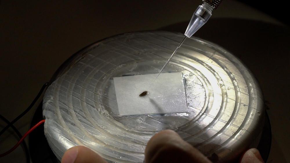
The new network was uncovered by testing many enzyme-encoding genes to narrow down which one facilitates signaling pathway crosstalk. The screening method works by inactivating, or silencing, RNA messages before they are translated into proteins. Then, following amputation, the team examined the effect of gene inactivation by measuring regeneration.
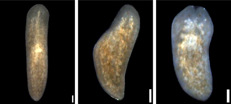
Silencing pak1 RNAs in flatworms with missing body parts resulted in some abnormal regeneration. Some regenerated a tail in place of a head, and others failed to readjust their middle-to-edge axis. These defects in regeneration indicate the importance of pak1 in mediating tissue regeneration along both axes.
The independent pathways that establish and maintain the head-to-tail and middle-to-edge axes have been previously described by the lab. How is then pak1 bridging these two pathways? The team hypothesized that pak1 likely functions with other genes to facilitate communication between the pathways. Researchers identified that pak1 works with a component of the Hippo pathway, merlin (mer), to bridge both the head-to-tail and middle-to-edge body axes.
Additional experiments revealed that pak1 and mer are both expressed in the same cells, leading the team to conclude that the enzyme (pak1) and signaling pathway for body size (Hippo) act together to coordinate the patterning and growth of new tissues during regeneration.
“I find it fascinating to investigate how cells read and process incoming information,” said Doddihal. “Our work is an attempt toward understanding the language of cells, providing a network for the flow of information and its processing during regeneration. We hope that our findings will help us advance our understanding of the fundamental principles of regeneration that restore biological form and function after injury.”
Additional authors include Frederick G. Mann, Jr., Ph.D., Eric Ross, Ph.D., Mary C. McKinney, Ph.D., Carlos Guerrero Hernández, Carolyn E. Brewster, and Sean A. McKinney, Ph.D.
This work was funded by the National Institutes of Health (NIH) (award: R37GM057260) and by institutional support from the Stowers Institute for Medical Research. The content is solely the responsibility of the authors and does not necessarily represent the official views of the NIH.