In The News

07 January 2026
Investigator Kamena Kostova, named ‘Cell Scientist to Watch’
From the Journal of Cell Science, Investigator Kamena Kostova named a 'Cell Scientist to Watch'
Read Article
News
Newly identified parental proteins present at fertilization reveal surprising levels of vertebrate developmental support

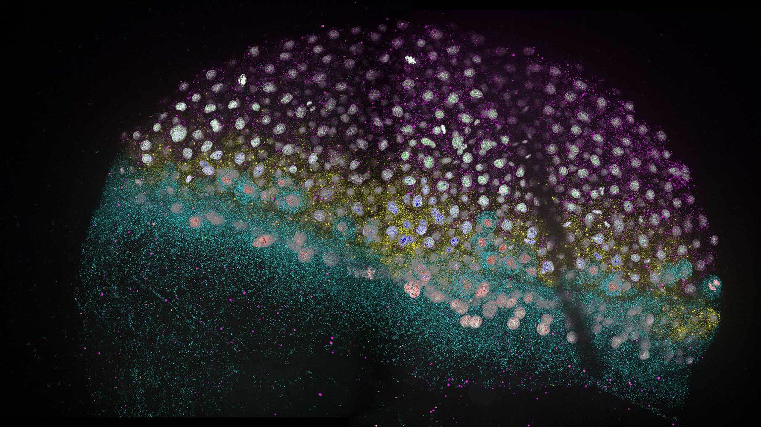
Fluorescent microscopy image showing zebrafish embryonic tissues (magenta, yellow, and cyan labeling) becoming specified early during development. Image courtesy of Cathy McKinney, Ph.D., from Microscopy and the Bazzini Lab.
By Rachel Scanza, Ph.D.
Mothers naturally shoulder the challenges of supporting the development of offspring during pregnancy. Now, new research from the Stowers Institute for Medical Research suggests that fathers may deserve recognition for contributions beyond half a genome.
From a single fertilized cell called a zygote, animals must undergo a critical transition during early embryogenesis. In this earliest stage of development, the zygote initially relies on molecules from the mother, already present in the egg cell, to activate its newly assembled genome and start making its own cellular components to grow.
A previous study from the lab of Stowers Institute Associate Investigator Ariel Bazzini, Ph.D., shed light on this transition, known as the maternal-to-zygotic transition by examining how the zebrafish zygote shifts from using maternally provided RNA messages toward manufacturing new RNAs. Now, a new study from the Bazzini Lab turns the focus from RNAs, many of which instruct the production of proteins, to the proteins themselves that are present during this critical transition in zebrafish. The study reveals new, surprising levels of developmental regulation.
“Support during the first moments of development for most animals is maternally skewed,” said Bazzini. “But our findings suggest that the father may provide proteins in addition to just half of the zygote’s genome, and those proteins might be key.”
The researchers were interested in exploring how gene expression, at the level of proteins, is regulated in an embryo after fertilization. The surprise was the identification of a set of genes known to be active later in development whose proteins are also deposited either maternally from the egg or paternally from the sperm at the time of fertilization, shared Gabriel da Silva Pescador, a predoctoral researcher in the Bazzini Lab.
“This study unravels newly identified processes at play during early embryogenesis that may translate toward a greater understanding of what happens early in human development,” said Bazzini.
Published in Cell Reports on September 19, 2024, the study led by da Silva Pescador in collaboration with Systems Mass Spectrometry, a Stowers Institute Technology Center, investigated fundamental processes during early development by characterizing proteins—their identities, concentrations, rates of accumulation, and stabilities—at specific time points spanning the maternal-to-zygotic transition.

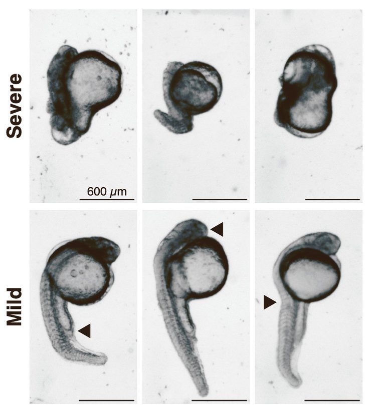
Microscopy images of severe and mild abnormalities observed in zebrafish embryos after CRISPR-Cas13d silencing of a critical developmental gene. Arrows point to tail shortening, deformation, or smaller head size.
“While the field of developmental biology typically focuses on RNA activity, we are showing that a full understanding of embryogenesis must take into account multiple regulatory levels—which RNAs are produced, the stability and concentration of these RNAs, how RNAs are translated into proteins, and the proteins themselves,” said Bazzini.
Combining RNA sequencing and previously published datasets of ribosome activity with a technique called protein profiling—a snapshot of all proteins expressed at a specific point in time—led to an unexpected finding. Of the over 2,000 proteins identified, 42 proteins were present at fertilization, but the RNA messages needed to produce them were not detected, suggesting that these proteins were already synthesized and deposited by either the sperm or egg.
The team distinguished these 42 proteins as coming from a previously identified “pure zygotic” gene pool, whose genes and corresponding proteins were thought to be exclusively produced by the zygote after its genome is fully functioning. Why protein products from pure zygotic genes were detected at fertilization puzzled the researchers but led them to hypothesize that these proteins may provide insight into another layer of regulation required during early embryogenesis.
“We found proteins potentially crucial for embryogenesis that we never would have detected if we had only looked at the RNA level,” said da Silva Pescador. “Actually, I reanalyzed the data three more times because conceptually, it was counterintuitive.”
To find the genetic source of these 42 proteins, the team combed through zebrafish sperm and egg RNA data. A few proteins could not be attributed to the egg’s RNAs but were found in sperm. Bazzini believes that using gene editing techniques to impair or remove genes that encode these proteins in sperm and egg cells should start to unveil the developmental function of these proteins.
“This research may impact our understanding of human fertility,” said da Silva Pescador. “If these newly identified maternal and paternal genes are not present, this could be one cause of human infertility with unknown origin.”
Along with uncovering newly identified maternally and paternally deposited proteins, the team used their protein profiling data to identify proteins that rapidly accumulate just before activation of the zygote’s genome. Using CRISPR-Cas13d, a gene editing technique for silencing RNAs that the Bazzini Lab previously optimized for animal embryos, the researchers targeted the RNA responsible for one protein whose concentration quickly increases. This allowed the identification of a novel gene that is indispensable for zebrafish development.
“We decided to explore this gene candidate further as other labs have identified how its human counterpart can disrupt cell fate in human cell lines when mutated,” said da Silva Pescador. “Silencing its RNA in zebrafish resulted in severe developmental abnormalities.”
This study marks the first time the technique has been used to identify a previously unknown function of a gene. In addition, the new protein profiling data set provides scientists with a long list of candidates that may be critical proteins for zygotic genome activation and can be evaluated using CRISPR-Cas13d.
Determining the processes that unfold during this brief but critical period after fertilization in zebrafish is important for understanding early development in vertebrates including humans.
“The study’s comprehensive analysis of protein dynamics during this transition could provide insight into early human development and potential causes of infertility that remain mysterious,” said da Silva Pescador.
Additional authors include Danielson Baia Amaral, Joseph Varberg, Ph.D., Ying Zhang, Ph.D., Yan Hao, and Laurence Florens, Ph.D.
This work was funded by the National Institute of General Medical Sciences of the National Institutes of Health (NIH) (award: GM136849), the NIH Office of the Director (award: R21OD034161), and through institutional support from the Stowers Institute for Medical Research. The content is solely the responsibility of the authors and does not necessarily represent the official views of the NIH.
In The News

07 January 2026
From the Journal of Cell Science, Investigator Kamena Kostova named a 'Cell Scientist to Watch'
Read Article
#Stowers25: Celebrating 25 Years
06 January 2026
Alejandro Sánchez Alvarado, Ph.D., reflects on a year of discovery, gratitude, and the community that helps support our mission.
Read Article
In The News

01 January 2026
From Science Friday, President and CSO Alejandro Sánchez Alvarado talks about the science of regeneration and the biology lessons we can carry into the new year.
Read Article
