News

09 October 2024
Road to Research: A Q&A with Laboratory Manager, Seth Malloy
"Every day presents a new challenge, a new question, and a new opportunity to positively impact the research community."
Read Article
The Histology team provides histotechnical instrumentation, technical support, and collaborative services to Stowers Institute scientists whose research involves the microscopic anatomy of biological tissues and cells.
Jump to:
Publications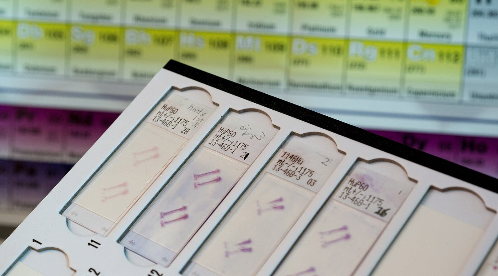

Comprised of histotechnologists and biologists with backgrounds in cytogenetics, molecular biology and neuroscience, the Histology Team collaborates with Stowers colleagues on projects to understand the spatial composition of RNA, protein, or other components in various types of samples, ranging from chromosome spread, cells, and tissue sections to whole mount organisms. The team has earned a reputation for creativity in developing histological methods to enable scientific discoveries.
The Stowers Histology center is a full-service histology laboratory that offers a variety of histological techniques to Institute members. We process fresh, paraffin-embedded and frozen tissue for routine histology, special stains, immunohistochemical assays, tissue clearing, in-situ hybridization, expansion microscopy, and spatial transcriptomics & proteomics. Additionally, we provide investigators with training, consultation and advice on how to best use histology services.
Team Contact
Director of Microscopy
Stowers Institute for Medical Research
Kexi Yi joined the Stowers Institute for Medical Research in 2009 as a research specialist in the lab of former Investigator Rong Li, Ph.D. He joined the Microscopy Technology Center in 2015 and was promoted to Head of Electron Microscopy in 2023.


Team Contact
Manager, Histology
Stowers Institute for Medical Research
Seth Malloy joined the Stowers Institute for Medical Research in the Histology Technology Center in 2018, and was promoted to manager of Histology in 2023.


News
09 October 2024
"Every day presents a new challenge, a new question, and a new opportunity to positively impact the research community."
Read Article
Press Release
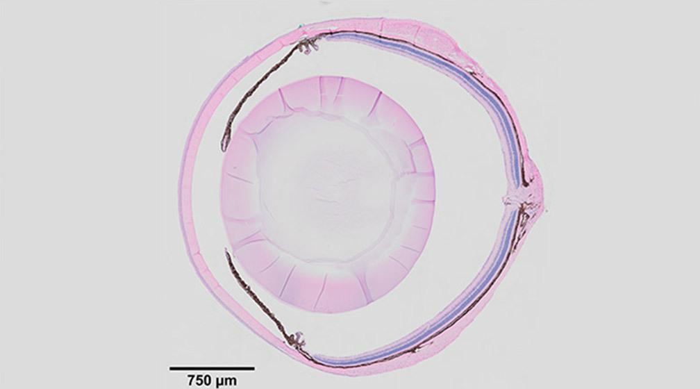
01 January 2021
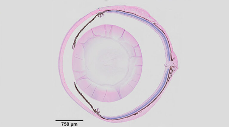
What if degenerative eye conditions could be detected and treated before vision is impaired? Recent findings from the Xie Lab point to the ciliary body as a key to unlocking this possibility.
Read Article
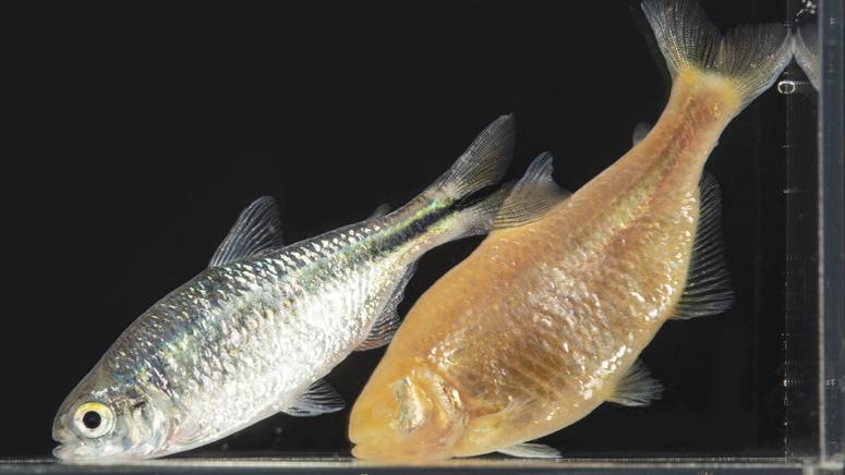
Video
04 September 2020
A comparative study of two fish species offers important clues about vertebrate regeneration.
Read Article
Pang J, Thomas N, Tsuchiya D, Parmely T, Yan D, Xie T, Wang Y. STAR Protoc. 2021;2:100879. doi: 100810.101016/j.xpro.102021.100879..
Changes in regeneration-responsive enhancers shape regenerative capacities in vertebrates
Wang W, Hu CK, Zeng A, Alegre D, Hu D, Gotting K, Ortega Granillo A, Wang Y, Robb S, Schnittker R, Zhang S, Alegre D, Li H, Ross E, Zhang N, Brunet A, Sanchez Alvarado A. Science. 2020;369: eaaz3090. doi: 10.1126/science.aaz3090.
Peuß R, Box AC, Chen S, Wang Y, Tsuchiya D, Persons JL, Kenzior A, Maldonado E, Krishnan J, Scharsack JP, Slaughter BD, Rohner N. Nat Ecol Evol. 2020;4:1416-1430.
Wang Y, Xu W, Maddera L, Tsuchiya D, Thomas N, Yu CR, Parmely T. J Histotechnol. 2019;42:193-201.
Wang Y, Yu Z, Cahoon CK, Parmely T, Thomas N, Unruh JR, Slaughter BD, Hawley RS. Nat Protoc. 2018;13:1869-1895.
Zeng A, Li H, Guo L, Gao X, McKinney S, Wang Y, Yu Z, Park J, Semerad C, Ross E, Cheng LC, Davies E, Lei K, Wang W, Perera A, Hall K, Peak A, Box A, Sánchez Alvarado A. Cell. 2018;173:1593-1608.e20.